Download
1 / 44
440 likes | 625 Views
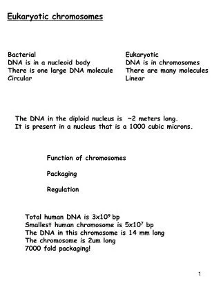
Eukaryotic chromosomes. Bacterial Eukaryotic DNA is in a nucleoid body DNA is in chromosomes There is one large DNA molecule There are many molecules Circular Linear. The DNA in the diploid nucleus is ~2 meters long. It is present in a nucleus that is a 1000 cubic microns.

E N D
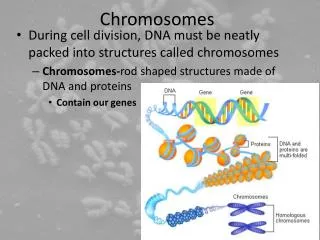
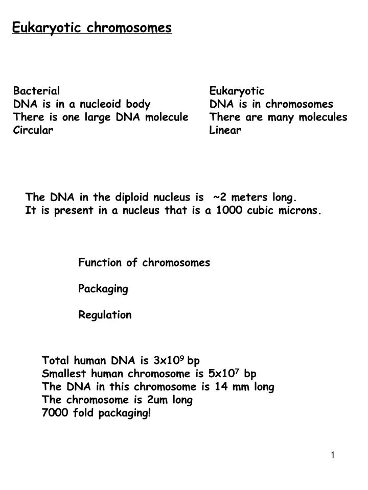
Eukaryotic chromosomes Bacterial Eukaryotic DNA is in a nucleoid body DNA is in chromosomes There is one large DNA molecule There are many molecules Circular Linear The DNA in the diploid nucleus is ~2 meters long. It is present in a nucleus that is a 1000 cubic microns. Function of chromosomes Packaging Regulation Total human DNA is 3x10⁹ bp Smallest human chromosome is 5x10⁷ bp The DNA in this chromosome is 14 mm long The chromosome is 2um long 7000 fold packaging!
Amount of DNA varies between species Amount of DNA varies in eukaryotes Salamander genomes are 20 times larger than human genomes Barley genome is 10 times larger than the rice genome Barley and rice are related. Measurements of DNA length Amount of DNA/nucleus = C value Species DNA content (pg or 10-12g) haploid Sponge 0.05 Drosophila 0.2 Human 3.5 Lungfish 102 Locust 46 Frog 4.2 Yeast 0.03
C-Value paradox This is often called the C value paradox. There is no phylogenetic relationship to DNA content There are sibling amphibian species - they look morphologically identical but have 4-fold difference in DNA content How do we account for the differences in DNA content/nucleus No of genes Gene size Distance between genes
Junk DNA 1) Number of genes could vary in these organisms Lungfish would have to have 30 fold more genes than humans Barley and rice have the same number of genes but vastly different DNA contents. Number of genes does not correlate with amount of DNA in a cell. 2) Size of genes could increase as genomes increase Drosophila genome is 30 times larger than E.coli Average coding region of a gene is 1-2 kb long in Drosophila E. Coli genes are only slightly shorter Drosophila genes are not 30 times larger than E. coli genes. Introns and promoters etc increase the size to some extent but cannot account for all of the increase. • 3) Amount of DNA between genes increases • Humans= 25,000 genes. Size of human genome is 3x10⁹bp • Yeast= 6000 genes. Size of yeast genome is 1.4x10⁷bp • The DNA between genes (intergenic region) varies. • A large fraction of intergenic DNA is repetitive • Nearly 60% of the human genome is repetitive. • Less than 5% of the yeast genome is repetitive.
Interphase nucleus Heterochromatin Inactive genes Euchromatin Gene Rich Active genes Constitutive heterochromatin Repetitive DNA (Telomere, centromere etc) Gene poor Transcriptionally silent Facultative heterochromatin Gene rich Transcriptionally silent
Epigenetics and development 2n DNA content same DNA content, > 200 cell types De-differentiation Differentiation • Cloning by nuclear transfer --> regenerate entire organism from transfer of single nucleus (e.g. Dolly) • Induced pluripotent stem cells (iPS) --> expression of 4 genes are sufficient to transform differentiated cells to “stem” cells • Both processes must involve reprogramming of epigenome!
Epigenetics: Gene regulation through stable activation/repression Heritable changes in gene activity that cannot be explained by changes in gene sequences This is essential for normal cell differentiation and development of an organism after fertilization Epigenetics imposes restrictions to the plasticity of totipotent embryonic cells During early development there is a progressive restriction of cellular plasticity accompanied by acquisition of cell type specific patterns of modifications on genes Epigenetic modifications impose a cellular memory that accompanies and enables stable differentiation
Epigenetic inheritance during mitosis sperm egg Embryo All Genes are poised for activity Cell commitment Specific genes activated All other genes inactivated Active genes maintain activity Inactive genes remain silent Active genes maintain activity Inactive genes remain silent
Epigenetics and gene ACTIVATION during development Heritable changes in gene expression that do not involve changes in DNA sequences All Genes not active in all cells • . examples: • Developmentally regulated / tissue specific gene expression • . mechanisms: • Presence of Transcription factors Active Inactive Transcription activator +++ ---
Gene activation TATA Inr Gene • examples: • Developmentally regulated / tissue specific gene expression • X chromosome dosage compensation • Gene Imprinting • Position effect variegation (PEV) Cell/tissue specific transcriptional activators bind to enhancers of genes that have binding sites for these factors -Aid in recruitment of enzymes that modify chromatin at the promoter - Aid in recruitment of the general transcription machinery and RNA polymerase Promoter Enhancer The enhancer functions to activate genes. There are specific sequences that bind TISSUE SPECIFIC transcription factors. The binding of these factors induces gene activation 100 fold! Active gene promoters have DNA sequences (CpG residues) that are not methylated and are bound by specific transcription activators.
Different Enhancers bind different tissue and cell specific transcription activator proteins and this enables specific gene activation in specific cells Cell specific Activation HNF3 Liver Cell Liver gene1 Brain gene1 HNF3 Liver gene2 Brain gene2 Brain Cell NZF2 Liver gene1 Brain gene1 NZF2 Liver gene2 Brain gene2
Epigenetics and gene REPRESSION during development Heritable changes in gene expression that do not involve changes in DNA sequences All Genes not active in all cells • . examples: • Developmentally regulated / tissue specific gene expression • . mechanisms: • Absence of transcription activators • Presence of repressors • Changes in Chromatin • Changes in DNA methylation Active Inactive Transcription activator +++ --- Repressor proteins --- +++ Specific Histone Modi +++ --- Specific Histone Modi --- +++ DNA methylation --- +++
sperm egg Embryo All Genes are poised for activity Cell commitment Specific genes activated All other genes inactivated Active genes maintain activity Inactive genes remain silent Active genes maintain activity Inactive genes remain silent • mechanisms: • Changes in Transcription factors • Changes in DNA methylation • Changes in Chromatin structure
Different Enhancers bind different tissue and cell specific transcription activator proteins and this enables specific gene activation in specific cells Cell specific Repression HNF3 Liver Cell Liver gene1 Brain gene1 HNF3 Liver gene2 Brain gene2 Brain Cell NZF2 Liver gene1 Brain gene1 NZF2 Liver gene2 Brain gene2
Gene Silencing and its importance In any given cell, only a small percentage of all genes are expressed Vast majority of the genome has to be shut down or silenced Knowing which genes to keep on and which ones to silence is critical for a cell to survive and proliferate normally during development and differentiation Transcription factors bind active genes and keep them active DNA methylation of inactive genes keeps them inactive Cell commitment Specific genes activated All other genes inactivated Active genes maintain activity Inactive genes remain silent
Inactive chromatin Heterochromatin Inactive Euchromatin Active • Constitutive heterochromatin: Repetitive DNA-Centromeres, telomeres etc • Repetitive DNA tends to recombine expanding/contracting repeats. Preventing repetitive DNA from recombination is critical for cell survival • Constitutes ~ 20 % of nuclear DNA • Highly compacted, • Always transcriptionally/Recombinationally inert • Euchromatin + facultative heterochromatin: • constitutes ~ 80% of nuclear DNA • less condensed, rich in genes, • Euchromatin is transcriptionally active • the rest is transcriptionally inactive (but can be activated in certain tissues or developmental stages) • These inactive regions are known as “facultative heterochromatin”
Facultative heterochromatin Regions of genome, rich in genes that are condensed in specific cell types or during specific stages of development. It includes genes that are highly active at a particular stage of development but then are stably repressed. X-chromosome inactivation in vertebrates (Dosage compensation) No. of transcripts are proportional to no. of gene copies Diploid- 2 copies of a gene Genes on X-chromosomes In females there are two copies of a gene. In males there is one copy. XX XY 2 1 Measuring transcript levels for genes on the X chromosome in female and male show that they are equivalent. Dosage imbalance is corrected!
examples: • Developmentally regulated / tissue specific gene expression • X chromosome dosage compensation • Gene Imprinting • Position effect variegation (PEV) Dosage compensation In Drosophila in the males there is an increase in transcription from the single X chromosome. A inhibitor of transcription is turned off in males allowing for full expression from the one X chromosome In nematodes there is a decrease in transcription from both X chromosomes- protein binds the 2X chromo and causes chromosome condensation which reduces transcription. In mammals, X chromosome inactivation occurs in females by formation of heterochromatin on one X chromosome
Mammalian X-chromosome inactivation Mammalian males and females have one and two X chromosomes respectively. One would expect that X-linked genes should produce twice as much gene product in females compared to males. Yet when one measures gene product from X-linked genes in males and females they are equivalent. This phenomenon, known as dosage compensation, X chromosome inactivation in females is the mechanism behind dosage compensation. In females, one of the X chromosomes in each cell is inactivated. This is observed cytologically. One of the X-chromosomes in females appears highly condensed. This inactivated chromosome is packaged into heterochromatin and forms a structure called a Barr-body.
Dosage compensation Dosage compensation in mammalian females occurs by shutting off of most of the genes on one X chromosome in females. The inactive X chromosome becomes heterochromatic. It is called a Barr body XCI is random. It occurs at the 500 cell stage of the embryo For a given cell in a developing organism, probability of the maternally or paternally derived X being inactivated is equal. Once inactivated, it is stably propagated so that all the thousands or millions of cells descended from that embryonic cell maintain the same chromosome in the Heterochromatic state. Xist is ON - Xist RNA coats the X- X chr is OFF Tsix is on- Tsix pairs and inactivates Xist -X chr is ON X chr with Xist gets methylated!!!! Genes on methylated X chromosome are not active.
XY XX reactivate X egg X sperm XX XX XX Tsix Active Xist Active Xist RNA Inactivates Xist RNA Coat inactive X- methylate DNA
CpG Methylation Epigenetic mechanism #1: DNA methylation • DNA methylation has long been correlated with repression of gene expression • DNA methylation mostly occurs on CpG dinucleotides and this modification is only observed on inactive gene promoters DNMTs methyl group added to the cytosine methylation status is maintained during replication/mitosis Methylated DNA recruits methyl-CpG-binding repressor proteins and other enzymes and this blocks access for RNA polymerase blocking transcription
X-inactivation The inactivation of one of the two X-chromosomes means that males and females each have one active X chromosome per cell. X-chromosome inactivation is random. For a given cell in the developing organism there is an equal probability of the female or the male derived X chromosome being inactivated.
X-inactivation zygote Inactivation Embryo The embryo is a mosaic! Once the decision is made in early development, then it is stably inherited. Patches of cells have the male X ON and patches of cells have the female X ON This is a Developmental rule that overlays on top of Mendellian rules!
Barr bodies · The inactive X-chromosome in normal females is called the barr body . XXX females have 2 Barr Bodies leaving one active X · XXXX females have 3 Barr Bodies leaving one active X · XXY males have one Barr Body leaving one active X (Klinefelter's syndrome) · X0 female have no Barr Bodies leaving one active X (Turner's syndrome) Given X-chromosome inactivation functions normally why are they phenotypically abnormal? Part of the explanation for the abnormal phenotypes is that the entire X is not inactivated during Barr-Body formation (Escape loci) Consequently an X0 individual is not genetically equivalent to an XX individual. XXY male XY male XX female XXX female
XmXf Xm Xm XmXf Xf Xf Xm Xm Xf Xf Mosaic expression XmXf XmXf XmXf XmXf XmXf XmXf XmXf XmXf XmXf
Tortoise shell cats Black Orange Enzyme O The O gene is carried on the X chromosome. Female cats heterozygous for the O gene on the X- chromosome have a particular pattern called Tortoise shell. According to Mendel’s rules the cats should be either orange or black. But the cats are neither! They are Tortoise shell.
All tortoiseshell cats are female XY male If normal OY gene is present on the X, the male is ginger If mutant oY gene is present in male it is black Female with O/O are ginger Females with o/o are black Females with O/o are tortoiseshell In O/o females X-chromosome inactivation happens at random Some cells activate O gene making ginger pigment Some cells activate o gene making black pigment Tortoiseshell cats
Tortoise shell cats According to Mendel’s rules these cats should be either orange or black. But the cats are neither! They are Tortoise shell. OO x oY F1 females are Oo
Tortoise shell cats Female cats heterozygous for the O gene on the X- chromosome have a particular pattern called Tortoise shell. According to Mendel’s rules these cats should be either orange or black. But the cats are neither! They are Tortoise shell.
DNA Methylation is not perfectly inherited during development/aging Fraga et al., 2005 PNAS 102(30):10604-9.
Epigenetic inheritance and meiosis sperm egg Embryo Active gene from father maintains activity Inactive gene from mother remain silent
examples: • Developmentally regulated / tissue specific gene expression • X chromosome dosage compensation • Gene Imprinting • Position effect variegation (PEV) Imprinting Occurs on Autosomes Occurs only on some genes on autosomes
Big bottom male x normal true breeding female 203 big bottom:209 normal C N N N C : N N N 50% 50% Normal male x big bottom female N N C N 100% normal Calliphyge is Sex independent- both males and females can be big bottom The callipyge gene is on autosome Big bottom is autosomal dominant?
CC x NN 100% Callipyge NN x CC 0% Callipyge Calliphyge gene is expressed when inherited from the males!!! The calliphyge locus from mother is always silenced.
* Callipyge Normal female XNormal male The callipyge locus from mother is always silenced. Normal phenotype female allele is imprinted (turned off) and male allele is expressed Normal female Xmutant male mutant female XNormal male * Mutant phenotype Normal allele (from mom) is imprinted (turned off) and mutant allele (from dad) is expressed Normal phenotype Mutant allele (from mom) is imprinted (turned off) and normal allele (from dad) is expressed
Imprinting A small number of genes (~200) on autosomes The allele from one parent is shut off. In the egg/sperm, these genes are imprinted (turned off) Imprinting leads to functional haploidy! Gene is WT but no protein is made (i.e. mutant). Abandoned safety net of diploidy. Gamete A=on A=off A=on Somatic cell A=off The original imprint is erased in gametes and the new imprint is established in progeny during gamete formation
DNA Methylation and imprinting CH3 CH3 Father IGF2 Gene Enhancer CTCF CTCF Mother IGF2 Gene Enhancer
War of the sexes Why are perfectly good genes turned off? Many maternally imprinted genes (inactive on the maternal chromosome) are fetal growth factor genes Tug of war Father contributes active genes to enhance growth- extract as many maternal resources for offspring as possible. He is unlikely to mate again with that female. Advantage for survival of his gene pool. Mother silences these growth promoting genes to ration her investment to any one offspring conserving resources for future.
The phenotype is expressed only when the mutant allele is inherited from the mother. Thus, mutant imprinted alleles can remain masked when they are paternally inherited, but clinically re-appear in one-half of children of carrier daughters