Download
1 / 106
1.22k likes | 1.94k Views
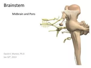
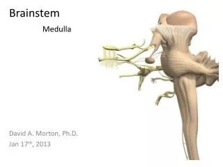
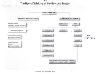
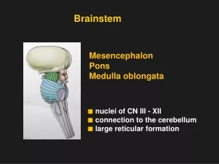
BRAINSTEM. Structural Overview of Brainstem. Midbrain, pons, medulla functions. BRAINSTEM. DORSAL SURFACE. VENTRAL SURFACE. medulla. Connects pons superiorly with spinal cord inferiorly Conical in shape

E N D
Structural Overviewof Brainstem • Midbrain, pons, medulla • functions
BRAINSTEM DORSAL SURFACE VENTRAL SURFACE
medulla • Connects pons superiorly with spinal cord inferiorly • Conical in shape • The Central canal continues upward into the lower half of medulla, in the upper half of medulla it expands as the cavity of fourth ventricle
two parts: open and closed • an open part (close to the pons) • a closed part (further down towards the spinal cord).
Gross Anatomy Review Medulla - ventral Anterior median fissure Pyramids Pyramidal decussation Olive Inferior cerebellar peduncle
Gross Anatomy Review Dorsal medulla • Posterior median sulcus • Gracile tubercle • Cuneate tubercle.
Brainstem X-sections • Caudal medulla • Rostral medulla • Caudal pons • Rostral pons • Caudal midbrain • Rostral midbrain
medulla • 3 sections inside the medulla: • Pyramidal decussation • Sensory decussation • Level of olive
CAUDAL MEDULLA(LEVEL OF PYRAMIDAL DECUSSATION) FG DMS FC GN ST5 Central grey matter CN SN5 Central canal DSC PD VSC P VMF
CAUDAL MEDULLA(LEVEL OF PYRAMIDAL DECUSSATION) • DMS: Dorsal median sulcus • FG: fasciculus gracilis • GN: Gracile nucleus • FC: Fasciculus cuneatus • CN: Cuneate nucleus • SN5: Spinal nucleus of trigeminal nerve • ST5: Spinal tract of trigeminal nerve • P: Pyramid • PD: Pyramidal decussation • DSC: Dorsal spinocerebellar tract • VSC: Ventral spinocerebellar tract • VMF: Ventral median fissure
CAUDAL MEDULLA(LEVEL OF PYRAMIDAL DECUSSATION) • GREY MATTER: • Sensory nuclei: Gracile, cuneate, spinal nucleus of trigeminal • WHITE MATTER: • Ascending tracts: Gracile, cuneate, spinal tract of trigeminal, dorsal & ventral spinocerebellar, spinal leminiscus • Descending tracts: Pyramidal & extrapyramidal tracts
CAUDAL MEDULLA(LEVEL OF PYRAMIDAL DECUSSATION) • Pyramidal decussation:Most of the fibers of pyramid decussate then pass laterally & dorsally to form the lateral corticospinal tract that descend in the lateral white column of spinal & terminate in ventral horn cells of opposite side • Spinal nucleus of trigeminal:It lies in the lower part of pons, the whole medulla & extends to the 2nd cervical segment of spinal cord where it is continuous with substantia gelatinosa. It receives pain & temperature sensations from the face along trigeminal nerve
CAUDAL MEDULLA(LEVEL OF PYRAMIDAL DECUSSATION) • Dorsal & ventral spinocerebellar tracts: They carry proprioceptive fibers to the cerebellum through inferior cerebellar peduncle (dorsal) & superior cerebellar peduncle (ventral) • Gracile &Cuneate tracts: They carry proprioceptive sensations & end in Gracile & Cuneate nuclei (2nd order neurones in dorsal column tract)
MID MEDULLA(LEVEL OF SENSORY DECUSSATION) DMS Central grey matter FG FC GN CN Central canal ST5 SN5 Internal Arcuate Fibers DSC M L VSC Sensory Decussation P VMF
MID MEDULLA(LEVEL OF SENSORY DECUSSATION) • Gracile & cuneate nuclei: They are more prominent. Axons of cells of gracile & cuneate nuclei curve around the central canal as internal arcuate fibers then decussate forming the sensory decussation & ascend in the brain stem as medial leminiscus that end in the ventral posterolateral nucleus of thalamus • Pyramid: They are more prominent
MID MEDULLA(LEVEL OF SENSORY DECUSSATION) • GREY MATTER: • Sensory nuclei: Gracile, cuneate, spinal nucleus of trigeminal • WHITE MATTER: • Ascending tracts: gracile, cuneate, spinal tract of trigeminal, dorsal & ventral spinocerebellar, spinal leminiscus • Descending tracts: Pyramidal & extrapyramidal tracts
Caudal medullaint arcuate fibers • gracile & cuneate nuc & fasc • Int arcuate fibers – ML • MLF • Nucleus of spinal tract of trigeminal nerve • Inferior olivary nuc • Pyramids • Hypoglossal nuclei
Rostral medullainf olivary nuc • olivary nuclear complex • Vestibulocochlear nuclei • Nucleus ambiguus • Hypoglossal nerve, dorsal nucleus of vagus, vestibulocochlear, glossopharyngeal and accessory nuclei
ROSTRAL MEDULLA DCN 4TH V MV LV V H S ICP VCN M L F A D Vagus Nerve I.O. ML M Hypoglossal Nerve P VMF
ROSTRAL MEDULLA • H: Hypoglossal nucleus • V: Dorsal vagal nucleus • S: Nucleus solitarius • A: nucleus ambiguus • MV: Medial vestibular nucleus • LV: Lateral vestibular nucleus • DCN: Dorsal cochlear nucleus • VCN: Ventral cochlear nucleus • ICP: Inferior cerebellar peduncle • I.O.: Inferior olive • D: Dorsal accessory olive • M: Medial accessory olive • MLF: Medial longitudinal fascisulus • ML: Medial leminiscus • P: Pyramid • VMF: Ventral median fissure
ROSTRAL MEDULLA • GREY MATTER: • Motor nuclei: Hypoglossal, dorsal vagal, nucleus ambiguus • Sensory nuclei: Nucleus solitarius, medial & lateral vestibular nuclei, dorsal & ventral cochlear nuclei, spinal nucleus of trigeminal • Extrapyramidal nuclei: Inferior olive, medial & dorsal accessory olive
ROSTRAL MEDULLA • WHITE MATTER: • Ascending tracts: Medial leminiscus, spinal leminiscus, spinal tract of trigeminal, ventral spinocerebellar tract • Descending tracts: Pyramidal & extrapyramidal tracts • Both ascending & descending tract: Medial longitudinal fasciculus • Inferior cerebellar peduncle: fibers connecting medulla to cerebellum
ROSTRAL MEDULLA • Hypoglossal nucleus: It lies in the medial part of floor of 4th ventricle. It contains motor neurones innervating muscles of tongue (except palatoglossus) through hypoglossal nerve • Dorsal vagal nucleus:It lies in the floor of 4th ventricle , lateral to hypoglossal nucleus. It contains preganglionic parasympathetic neurones running in the vagus nerve • Nucleus Solitarius:It lies ventrolateral to dorsal vagal nucleus. It receive taste fibers from facial, glossopharyngeal & vagus nerves
ROSTRAL MEDULLA • Nucleus ambiguus:It lies dorsal to inferior olivary nucleus. It contains motor neurones innervating muscles of pharynx, palate & larynx through glossopharyngeal, vagus & cranial part of accessory nerves • Medial & lateral vestibular nuclei:They lie in the floor of 4th ventricle, lateral to dorsal vagal nucleus. They receive afferent fibers from vestibular nerve • Dorsal & ventral cochlear nuclei:They lie dorsal (dorsal nucleus) & lateral (ventral nucleus) to ICP. They receive afferent fibers from cochlear nerve
ROSTRAL MEDULLA • Olivary nuclear complex:It is formed of a large nucleus (inferior olive) & 2 smaller nuclei (medial & dorsal accessory olive). • Afferents: From cerebral and cerebellar cortex & spinal cord • Efferents: To cerebellum through ICP • Function: They are concerned with control of movement
ROSTRAL MEDULLA • Medial longitudinal fasciculus:It consists of both ascending & descending fibers: • Ascending fibers: connect vestibular nuclei to nuclei supplying extraoccular muscles (occulomotor, trochlear & abducent nuclei). It coordinates movements of head & eyes • Descending fibers: connect vestibular nuclei to nuclei of ventral horn of spinal cord (medial vestibulospinal tract). It control body posture & balance
ROSTRAL MEDULLA • Spinal leminiscus: It carries pain, temperature & touch sensations from the opposite side of body to ventral posterolateral nucleus of thalamus • Inferior cerebellar peduncle: It is formed of fibers connecting medulla to cerebellum
Level of Inferior Olives Vestibular nuclei Medial Inferior Hypoglossal nucleus CN XII Inferior cerebellar peduncle = Restiform body Inferior olivary nuclei MLF Arcuate nuclei pontine nuclei
Rostral medulla N. solitarious Sensory nucleus for CN VII, IX, X Dorsal motor nucleus of X Spinal trigeminal tract CN V, VII, IX, X N. ambiguus Motor nucleus for CN IX, X & XI
Cranial Nerves of the Medulla Vestibular nuclei CN XII
Cranial Nuclei of the Medulla N. solitarious Sensory nucleus for CN VII, IX, X
Cranial Nuclei of the Medulla N. solitarious Sensory nucleus for CN VII, IX, X Spinal trigeminal tract
Cranial Nuclei of the Medulla N. solitarious Sensory nucleus for CN VII, IX, X Spinal trigeminal tract N. ambiguus Motor nucleus for CN IX, X & XI
Cranial Nuclei of the Medulla N. solitarious Sensory nucleus for CN VII, IX, X Dorsal motor nucleus of X Spinal trigeminal tract CN V, VII, IX, X N. ambiguus Motor nucleus for CN IX, X & XI
CN IX: Glossopharyngeal Nerve N. solitarious Sensory nucleus for CN VII, IX, X Spinal trigeminal tract CN V, VII, IX, X N. ambiguus Motor nucleus for CN IX, X & XI
CN IX: Glossopharyngeal Nerve N. solitarious Sensory nucleus for CN VII, IX, X Posterior 1/3 of the tongue Spinal trigeminal tract CN V, VII, IX, X N. ambiguus Motor nucleus for CN IX, X & XI
CN IX: Glossopharyngeal Nerve N. solitarious Sensory nucleus for CN VII, IX, X Posterior 1/3 of the tongue Spinal trigeminal tract CN V, VII, IX, X Sensation behind ear N. ambiguus Motor nucleus for CN IX, X & XI
CN IX: Glossopharyngeal Nerve N. solitarious Sensory nucleus for CN VII, IX, X Posterior 1/3 of the tongue Spinal trigeminal tract CN V, VII, IX, X Sensation behind ear N. ambiguus Motor nucleus for CN IX, X & XI Stylopharyngeus (lifts pharynx)
CN IX: Glossopharyngeal Nerve N. solitarious Sensory nucleus for CN VII, IX, X Posterior 1/3 of the tongue Inf. salivatory nucleus Parotid gland, parasympathetic Spinal trigeminal tract CN V, VII, IX, X Sensation behind ear N. ambiguus Motor nucleus for CN IX, X & XI Stylopharyngeus (lifts pharynx)
CN X: Vagus Nerve N. solitarious Sensory nucleus for CN VII, IX, X Spinal trigeminal tract CN V, VII, IX, X N. ambiguus Motor nucleus for CN IX, X & XI
CN X: Vagus Nerve N. solitarious Sensory nucleus for CN VII, IX, X Taste, epiglottis Cardiorespiratory Spinal trigeminal tract CN V, VII, IX, X N. ambiguus Motor nucleus for CN IX, X & XI
CN X: Vagus Nerve N. solitarious Sensory nucleus for CN VII, IX, X Taste, epiglottis Cardiorespiratory Spinal trigeminal tract CN V, VII, IX, X Ear N. ambiguus Motor nucleus for CN IX, X & XI
CN X: Vagus Nerve N. solitarious Sensory nucleus for CN VII, IX, X Taste, epiglottis Cardiorespiratory Spinal trigeminal tract CN V, VII, IX, X Ear N. ambiguus Motor nucleus for CN IX, X & XI Pharynx Larynx
CN X: Vagus Nerve N. solitarious Sensory nucleus for CN VII, IX, X Taste, epiglottis Cardiorespiratory Dorsal motor nucleus of X Spinal trigeminal tract CN V, VII, IX, X Ear N. ambiguus Motor nucleus for CN IX, X & XI Pharynx Larynx