Download
1 / 28
360 likes | 886 Views
Brainstem. Assoc. Prof. Stefan Sivkov, Ph.D. Dept. of Anatomy, Histology and Embryology. Development. Ventricles in brainstem. Mesencephalon cerebral aqueduct Metencephalon 4 th ventricle Mylencephalon 4 th ventricle. Posterior commissure. Corpus callosum. Fornix.

E N D
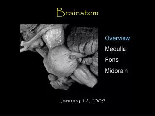
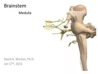
Brainstem Assoc. Prof. Stefan Sivkov, Ph.D. Dept. of Anatomy, Histology and Embryology
Ventricles in brainstem • Mesencephalon cerebral aqueduct • Metencephalon 4th ventricle • Mylencephalon 4th ventricle
Posterior commissure Corpus callosum Fornix Occipital Lobe Thalamus Anterior commissure Quadrigeminal cistern Hypothalamus vermis Optic nerve 4th ventricle Mammillary body pyramid
basal ganglia internal capsule optic chiasm optic nerve optic tract hypothalamus mammillary body cerebral peduncle pons interpeduncular fossa flocculus inferior olivary nuclear complex cerebellar tonsil cerebellum pyramidal decussation pyramid Anterior view of brainstem
cerebral peduncle optic tract trigeminal nerve optic nerve middle cerebellar peduncle optic chiasm vestibulocochlear nerve flocculus hypothalamus cuneate tubercle pons inferior olivary nuclear complex pyramid anterior median fissure Lateral view of brainstem
Posterior view of brainstem Superior colliculus Cerebral peduncle Inferior colliculus Superior cerebellar peduncle 4th ventricle Middle cerebellar peduncle Inferior cerebellar peduncle Medulla
Components of the brainstem • Sensory ascending pathways (dorsal): • Relay nuclei, tracts • Motor descending pathways (ventral) • Tracts, motor nuclei brainstem • Cerebellar pathways • Tracts, cerebellar afferent and efferent nuclei • Cranial nerve sensory and motor tracts • Cranial nerve nuclei, nerve entry and exit points • CPGs: rhythmic chewing, respiration, cardiovascular regulation & gain adjustments for reflexes • Modulatory systems: locus coeruleus, raphe & substantia nigra • Chemically coded nuclei
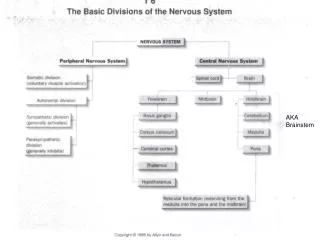
Brain Stem • Located btwn the cerebrum and the SC • Provides a pathway for tracts running btwn higher and lower neural centers. • Consists of themidbrain, pons, andmedullaoblongata. • Each region is about an inch in length. • Microscopically, it consists of deep gray matter surrounded by white matter fiber tracts. • Produce automatic behaviors necessary for survival.
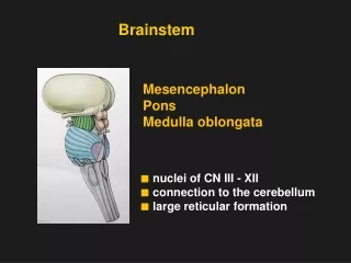
Brainstem: 3 major divisions • Midbrain • Pons • Medulla
Midbrain • Located btwn diencephalon and pons. • 2 bulgingcerebral peduncleson the ventral side. These contain: • Descending fibers that go to the cerebellum via the pons • Descending pyramidal tracts • Running thru the midbrain is the hollowcerebral aqueductwhich connects the 3rd and 4th ventricles of the brain. • The roof of the aqueduct (thetectum) contains thecorpora quadrigemina • 2superior colliculithat control reflex movements of the eyes, head and neck in response to visual stimuli • 2inferior colliculithat control reflex movements of the head, neck, and trunk in response to auditory stimuli
Cranial nerves 3&4 (oculomotor and trochlear) exit from the midbrain • Midbrain also contains the headquarters of the reticular activating system
Midbrain • On each side, the midbrain contains ared nucleusand asubstantia nigra • Red nucleus contains numerous blood vessels and receives info from the cerebrum and cerebellum and issues subconscious motor commands concerned w/ muscle tone & posture • Lateral to the red nucleus is the melanin-containing substantia nigra which secretes dopamine to inhibit the excitatoryneurons of the basal nuclei. • Damage to the substantia nigra would cause what?
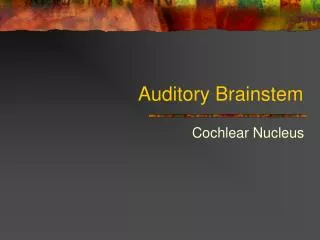
Pons • Literally means “bridge” • Wedged btwn the midbrain & medulla. • Contains: • Sensory and motor nuclei for 4 cranial nerves • Trigeminal (5), Abducens (6), Facial (7), and Auditory/Vestibular (8) • Respiratory nuclei: • Apneustic&pneumotaxic centerswork w/ the medulla to maintain respiratory rhythm • Nuclei & tracts that process and relay info to/from the cerebellum • Ascending, descending, and transverse tracts that interconnect other portions of the CNS
Medulla Oblongata • Most inferior region of the brain stem. • Becomes the spinal cord at the level of the foramen magnum. • Ventrally, 2 ridges (themedullary pyramids)are visible. • These are formed by the large motorcorticospinal tracts. • Right above the medulla-SC junction, most of these fibers cross-over (decussate).
Medulla Oblongata • Nuclei in the medulla are associated w/ autonomic control, cranial nerves, and motor/sensory relay. • Autonomic nuclei: • Cardiovascular centers • Alter the rate and force of cardiac contractions • Alter the tone of vascular smooth muscle • Respiratory rhythmicity centers • Receive input from the pons • Additional Centers • Emesis, deglutition,coughing, hiccupping, and sneezing
Medulla Oblongata • Sensory & motor nuclei of 5 cranial nerves: • Auditory/Vestibular (8), Glossopharyngeal (9), Vagus (10), Accessory (11), and Hypoglossal (12) • Relay nuclei • Nucleus gracilisandnucleus cuneatuspass somatic sensory information to the thalamus • Olivary nucleirelay info from the spinal cord, cerebral cortex, and the brainstem to the cerebellar cortex.
Ascending sensory pathways Fine discriminitive touch, conscious proprioception • Fasciculus gracilis: Terminates in the nucleus gracilis (medulla) • Fasciculus cuneatus: Terminates (medulla) in the cuneate and accessory cuneate nuclei Sensations of pain and temperature • Lateral Spinothalamic Tract • origin dorsal horn cells of the gray matter • Fibers cross contralaterally through the anterior commissure and ascend to the VPL nucleus Transmits sensations of touch • Ventral Spinothalamic Tract • origin cells of the posterior horn • Fibers cross to the opposite side in the anterior commissure
Descending motor pathways Voluntary movement • Lateral Corticospinal Tract • Originates in large pyramidal cells (precentral gyrus) • cross to the opposite side of the cord at the pyramidal decussation & terminate in the dorsal horn cells • Ventral Corticospinal Tract • Originates in the pyramidal cells (motor area of the cortex) Impulses related to equilibrium and antigravity reflexes • Vestibulospinal Tract • Fibers originate in the vestibular nuclei of the medulla and terminate at level of the sacral spinal nerves Connects vestibular complex and head and eye movement coordination center in medulla • Medial Longitudinal Fasciculus • Contains both ascending and descending fibers
Motor Hierarchy • Lateral group (extremities; fine motor control) • Corticospinal tract • Rubrospinal tract • Medial group (axial musculature; rhythmic and postural movements) • Vestibulospinal tract • Tectospinal tract • Reticulospinal tract • “Final common path”: motor pool
Reticular Formation • Extensive network of neurons that runs thru the medulla and projects to thalamic nuclei that influence large areas of the cerebral cortex. • Midbrain portion of RAS most likely is its center • Functions as a net or filter for sensory input. • Filter out repetitive stimuli. Such as? • Allows passage of infrequent or important stimuli to reach the cerebral cortex. • Unless inhibited by other brain regions, it activates the cerebral cortex – keeping it alert and awake. How might the “sleep centers” of your brain work? Why does alcohol make you tired?
Reticular Formation • “Core” of brainstem (midbrain, pons and medulla) composed of loosely organized neurons, outside of the major nuclear groups of the brainstem. • Medial-to-lateral: raphe nuclei, gigantocellular region, small cell region • Participate in widespread connections • Rostral continuation of interneuronal network found in spinal cord
Dorsal Column/Medial Lemniscal system • Secondary neuron is in brainstem: nucleus gracilis and cuneatus=dorsal column nuclei • Output of dorsal column nuclei crosses midline and forms recognizable bundle: medial lemniscus • Medial lemniscus fibers synapse in the thalamus in the ventroposterior nuclei • Thalamic axons synapse in primary somatosensory cortex in several somatotopic maps with some segregation of submodalities http://cas.bellarmine.edu/tietjen/HumanBioogy/central_nervous_system.
Tracing through the brainstem: Dorsal Column/Medial Lemniscal System