Download
1 / 35
440 likes | 833 Views
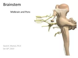
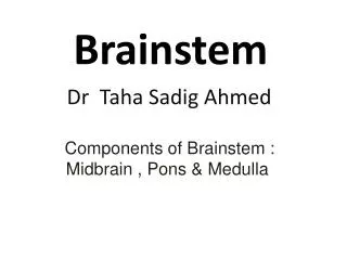
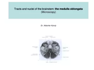
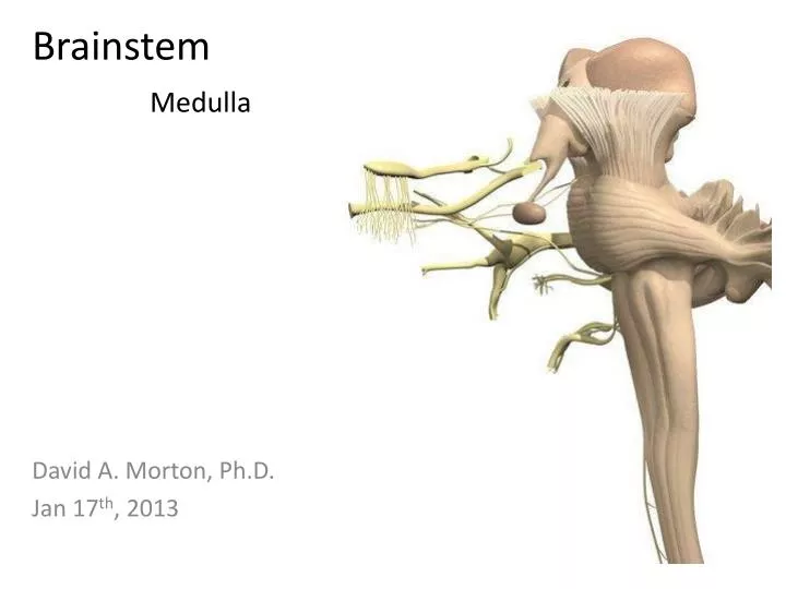
Brainstem Medulla. David A. Morton, Ph.D. Jan 17 th , 2013. Objectives. Describe the trajectory of the cranial nerves, their components, and their functions Identify and locate the CN’s associated with the medulla, the pons and the midbrain.

E N D
BrainstemMedulla David A. Morton, Ph.D. Jan 17th, 2013
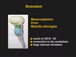
Objectives • Describe the trajectory of the cranial nerves, their components, and their functions • Identify and locate the CN’s associated with the medulla, the pons and the midbrain. • Explain how cranial nerves differ from spinal nerves • List the cranial nerves that contain parasympathetic fibers, the location of their nuclei, and their function • Recognize the major internal and external landmarks on the dorsal and ventral surface of the brain stem, so that you can determine if a gross or stained cross section is medulla, pons or midbrain. • Identify on a typical cross section all the brain stem nuclei containing motor neurons that end on striated muscle. • Explain why cranial nerves are so important in localizing lesions. • Name reflexes that test these nerves and brain stem levels. • Relate branches of the vertebrobasilar blood supply to the medulla and pons explaining the deficits that would occur with vascular occlusion.
Internal anatomy of brainstem • The fate of the alar and basal laminae • Why are brain stem sensory nuclei lateral to motor nuclei in brainstem? Sulcus limitans Som S Som S Alar Alar VS VS VM Sulcus limitans SM BM VM Basal SM Basal Medulla Spinal cord
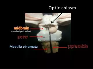
Medulla oblongata • External anatomy: • Pyramid • Olive • CNN VIII, IX, X, XI and XII • Fourth ventricle • Vertebral arteries 4th vent. VIII P O IX & X Medulla XII XI O P Horizontal section
Medulla oblongata Solitary nuclei and tract Spinal trigeminal nucleus Inf sal and dorsal vagal nuclei Sulcus limitans Hypoglossal nucleus Nucleus ambiguus Som S Alar VS VM SM BM Basal Medulla
Medulla Oblongata • Hypoglossal nucleus (CN XII) • Somatic motor • Comparable to ventral horn • Tongue muscles
Medulla Oblongata • Dorsal motor nucleus (CN X) • Visceral motor • Comparable to lateral horn • Origin of preganglionic parasympathetic neurons
Medulla Oblongata • Inferior salivatory nucleus (CN IX) • Visceral motor • Comparable to lateral horn (can not see it on brains) • Origin of preganglionic parasympathetic neurons to otic ganglion
Medulla Oblongata • Nucleus ambiguus (CN IX and X) • Branchial (spec visc) motor • Origin of BM neurons for IX and X
Medulla Oblongata • Reticular formation • Forms the central core of brain stem • Nuclear groups not obvious
Medulla Oblongata • Vascular supply: • Vertebra artery • Anterior spinal artery • PICA PICA Vert Ant sp
Objectives • Explain what the meninges cover and what spaces they surround. • For each meningeal space describe a classic source for blood in the space. • Describe where CSF is produced and how it circulates and is removed. • Name the most likely sites of obstruction of CSF circulation and the consequences. • Explain how the Blood Brain Barrier is different from the CSF Brain interface.
Cranial Meninges Dura mater Arachnoid mater Pia mater
Cranial Meninges • Dura mater (2 layers in the skull) • Periosteal layer and Meningeal layer • Dural venous sinus Dural Venous sinuses Cerebral vv.
Meningeal hemorrhages • Blood in meningeal spaces or potential spaces • Epidural hemorrhage • Subdural hemorrhage • Subarachnoid hemorrhage
Epidural space • Dura mater • Middle meningeal artery
Epidural hemorrhage Torn middle meningeal a.
Subdural space • Dura mater • Bridging cerebral vein • Courses between cerebrum and dural venous sinus
Torn bridging cerebral v. Subdural hemorrhage
What space is the artery located? Dural Venous sinuses
Brain arteries – Subarachnoid space • Internal carotid artery (ICA) • Ophthalmic • Anterior cerebral • Middle cerebral • Vertebral artery • PICA • Basilar • AICA • SCA • Posterior cerebral • Cerebral arterial circle of Willis • Anterior communicating • Anterior cerebral • ICA • Posterior communicating • Posterior cerebral
Organize the following terms: Cerebral aqueduct Lateral ventricle 4th ventricle Arachnoid granulations Superior sagittal sinus Lateral apertures 3rd ventricle Subarachnoid space Aqueduct of Sylvius Interventricular foramen Foramen of Magendie Median aperture Foramen of Monro Foramen of Luschka Choroid plexus Lateral ventricle Mesencephalic aqueduct
The following terms are in order: Choroid plexus Lateral ventricle Lateral ventricle Interventricular foramen Foramen of Monro 3rd ventricle Cerebral aqueduct Aqueduct of Sylvius Mesencephalic aqueduct 4th ventricle Lateral apertures Median aperture Foramen of Luschka Foramen of Magendie Subarachnoid space Arachnoid granulations Superior sagittal sinus
The ventricular system The ventricles, lined by ependymal cells, are obvious internal landmarks, and are important as internal structures to relate other structures to. Try to visualize them and rotate them with the brain. They will form the “wire frame” for our orientation to the brain.
The ventricular system • Hydrocephalus is dilation of all or part of the ventricular system due to obstruction of CSF flow. There are two types: • 1. Obstructive (non-communicating) hydrocephalus. Block in the ventricular system or the outlet foramina. • 2. Communicating (non-obstructive) hydrocephalus. Block in the subarachnoid space or the arachnoid granulations. • a. Meningitis followed by scarring. • b. Subarachnoid hemorrhage, meningeal fibrosis and scarring
Hydrocephaly 1. Obstructive (non-communicating) hydrocephalus. Block in the ventricular system or the outlet foramina.
2. Communicating (non-obstructive) hydrocephalus. Block in the subarachnoid space or the arachnoid granulations. Hydrocephaly
Figure 12.26a Formation, location, and circulation of CSF. Superior sagittal sinus 4 Choroid plexus Arachnoid villus Interventricular foramen Subarachnoid space Arachnoid mater Meningeal dura mater Periosteal dura mater 1 Right lateral ventricle (deep to cut) Choroid plexus of fourth ventricle 3 Third ventricle 1 CSF is produced by the choroid plexus of each ventricle. Cerebral aqueduct Lateral aperture 2 CSF flows through the ventricles and into the subarachnoid space via the median and lateral apertures. Some CSF flows through the central canal of the spinal cord. Fourth ventricle Median aperture 2 Central canal of spinal cord 3 CSF flows through the subarachnoid space. (a) CSF circulation 4 CSF is absorbed into the dural venous sinuses via the arachnoid villi.