Download
1 / 35
460 likes | 1.35k Views
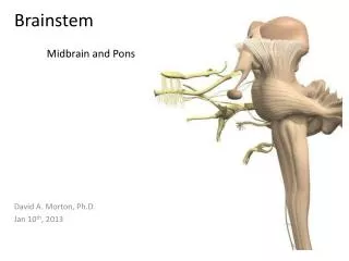
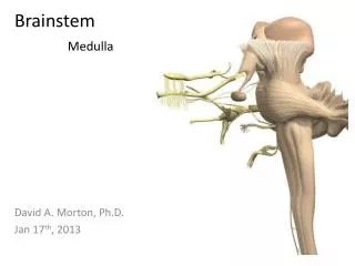
Brainstem II. Medical Neuroscience Dr. Wiegand. Internal Brainstem. Cranial nerve nuclei Location of selected tracts Reticular formation. Developmental Organization. Developmental Organization. Sulcus Limitans. Developmental Organization. From Pritchard & Alloway: Fig. 4-1.

E N D
Brainstem II Medical Neuroscience Dr. Wiegand
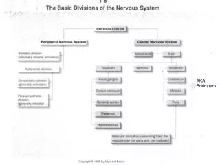
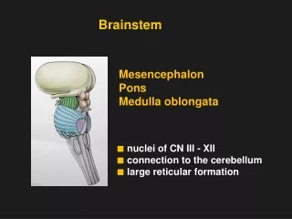
Internal Brainstem • Cranial nerve nuclei • Location of selected tracts • Reticular formation
Developmental Organization Sulcus Limitans
Developmental Organization From Pritchard & Alloway: Fig. 4-1
From Pritchard & Alloway: Fig. 4-4 SEN MOT Cranial Nerve Nuclei Organization • Medial to sulcus limitans • GSE SVE GVE • Lateral from sulcus limitans • VA GSA SSA
Generalizations • Sensory nuclei lateral to sulcus limitans • Motor nuclei medial to sulcus limitans • Visceral nuclei are on either side of sulcus • Innervation of skeletal muscle (GSE & SVE) most medial • General and special visceral afferent nuclei in same column
I, II Cranial Nerves – Telencephalon & Diencephalon • Olfactory – • smell (SVA) • Optic – • vision (SSA)
III, IV Cranial Nerves –Mesencephalon • Oculomotor – • extraocular eye muscles (GSE) – oculomotor nucleus • PSNS to eye (GVE) – Edinger-Westphal nucleus • Trochlear – • extraocular muscle (sup. oblique) (GSE) – trochlear nucleus
V, VI Cranial Nerves –Metencephalon • Trigeminal – • Masticatory muscles (SVE) – trigeminal motor nucleus • General sensation of the head and face (GSA) – trigeminal complex • Abducens – • extraocular muscle (lat. rectus) (GSE) – abducens nucleus
VII Cranial Nerves –Metencephalon • Facial – • Facial expression muscles (SVE) – facial motor nucleus • Glands (submandibular, sublingual & lacrimal) (GVE) – superior salivatory & lacrimal nucleus • Taste (SVA) – rostral solitary nucleus • General sensation of ear (GSA) – trigeminal complex
VIII Cranial Nerves –Metencephalon Vestibulocochlear – • Hearing (SSA) – dorsal and ventral cochlear nuclei • Balance (SSA) – vestibular nuclei
IX Cranial Nerves –Mylencephalon • Glossopharyngeal • Stylopharyngeus muscle (SVE) – n. ambiguus • PSNS to parotid gland (GVE) – inferior salivatory n. • Taste (SVA) – rostral solitary n. • Carotid body sensation (GVA) – caudal solitary n. • General sensation from ear & tongue (GSA) – trigeminal complex
IX Cranial Nerves –Mylencephalon • Vagus • Muscles of larynx & pharynx (SVE) – n. ambiguus • PSNS to thorax and upper abdomen (GVE) – dorsal motor n. of X (DMV) • Sensory from viscera (GVA) – caudal solitary n. • Taste (SVA) – rostral solitary n. • General sensation from ear (GSA) - trigeminal complex
XI, XII Cranial Nerves –Mylencephalon • Accessory – • innervates trapezius and sternocleidomastoid (SVE or GSE) – motor nucleus of XI in upper cervical cord • Hypoglossal – • tongue muscles (GSE) – hypoglossal nucleus
Cranial Nerve Nuclei Groups • GSE & SVE – Motor Nuclei of: • III, IV, V, VI, VII, XI, XII • ambiguus • GVE – Parasympathetic Nuclei • Edinger-Westphal nucleus • Lacrimal & salivatory (superior, inferior) nuclei • Dorsal Motor Nucleus of X • SVA (Taste) – Rostral Solitary nucleus • GVA – Caudal Solitary nucleus • GSA – Trigeminal (TBNC) • SSA – Cochlear and Vestibular nuclei
http://lansing.bellarmine.edu/pt/atlas/cover.html Learning Internal Anatomy • Recognize outline of brainstem • Midbrain • Inferior vs. superior colliculi • Pons • Medulla • Open vs. closed • Place nuclei in correct level • Recognize orientation of slice • Learn Pathways • Relationship of tracts • Places of decussation
Sensory Nuclei Sensory Decussation Medial Lemniscus Anterolateral System Spinocerebellar Tracts ASCENDING PATHWAYS
Corticospinal & Corticobulbar Tracts (Pyramidal Tract) DESCENDING PATHWAY
Edinger-Westphal Nuclei Oculomotor Nuclei Trigeminal Nerve Trigeminal Nuclei Hypoglossal Nucleus Dorsal Motor of Vagus Solitary Nucleus CRANIAL NERVES
Inferior olivary nucleus Red nucleus Substantia Nigra Other Brainstem Nuclei
Reticular Formation • Diffuse, poorly differentiated brainstem nuclei • Occupies tegmentum of brainstem • Modulates: • Pain • Muscle tone and reflexes • Autonomic functions e.g. respiration, blood pressure, cardiac function • Arousal, awareness and attention
Reticular Formation Descending Input Descending Input Forebrain & Brainstem Nuclei Cerebellar projecting nuclei Cerebellar projecting nuclei Raphe lateral medial medial lateral Spinal Cord & Brainstem Nuclei Ascending Input Ascending Input
Midline Raphe Nuclei • Mostly serotoninergic cells – send and receive extensive ascending and descending projections • Regulate pain, arousal and sleep
Parasagittal medial nuclei • Motor regulation • Medial (facilitates extensors) reticulospinal tract • Lateral (facilitates flexors) reticulospinal tract • Also autonomic regulation • ARAS exerts excitatory input to cortex for consciousness and attention
Locus Ceruleus & Medial Reticular Zone • Regulates attention • Inhibits pain at spinal cord level • Regulates autonomic function
Parasagittal Lateral Nuclei • Receives input to mediate visceral and cranial nerve reflexes • Projects to parasagittal medial nuclei • Pedunculopontine n.
Pedunculopontine Nucleus • Acetylcholine projecting cells • Ascending projection to inferior frontal cortex/intralaminar n. • Input from basal ganglia output nuclei • Projects to brainstem motor nuclei (VST, RST)
Wallenberg’s Syndrome Lateral medullary syndrome Thrombosis of PICA
Wallenberg’s Syndrome • Structures involved: • Inf. Cerebellar peduncle (ipsilateral ataxia) • SpV tract & nucleus (ipsilateral loss pain & temperature) • Anterolateral system (contralateral loss of body pain & temperature) • N. ambiguus (dysphagia & dysphonia) • Vestibular n. (nystagmus and postural instability)