Download
1 / 30
400 likes | 843 Views
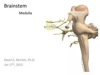
Brainstem 3. Midbrain. Dr Rania Gabr. Objectives. Identify the gross features of the brainstem.

E N D
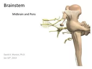
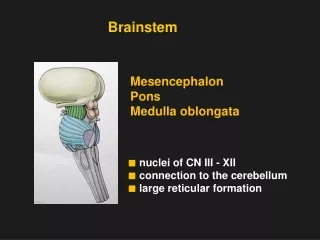
Brainstem 3 Midbrain Dr Rania Gabr
Objectives • Identify the gross features of the brainstem. • Briefly describe the internal structure of the brainstems (ascending and descending pathways, sensory and motor cranial nuclei, substantianigra, red nucleus, olivary nucleus and reticular formation). • Describe the main connections of the sensory cranial nuclei. • Describe the main connections of the motor cranial nuclei. • Review the blood supply of the brainstem. • Describe lesions in the brainstem such as medial medullary syndrome and lateral medullary syndrome. • Describe the main connections of the substantianigra and the red nucleus.
Midbrain Cruscerebri • Shortest part of the brain stem, not more than 2cm in length, lies in the posterior cranial Fossa. • For descriptive purposes, divided into: • Dorsal tectumand right and left Cerebral Peduncles. • -Cerebral peduncles contains: • 1-Descending fibers that go to the cerebellum via the pons • 2-Descending pyramidal tracts • -Running through the midbrain is the hollow cerebral aqueduct which connects the 3rd and 4th ventricles of the brain.
Each cerebral peduncle divides further into ventralcrus cerebri (massive fibrous mass)and a dorsalTegmentumby a pigmented lamina “ Substantianigra”
MID BRAIN – VENTRAL SURFACE • Large column of descending fibers (crus cerebri or basis pedunculi), on either side, separated by a depression called the interpeduncular fossa with posterior perforated substance. • Nerve emerging from Midbrain (one): • Occulomotor (3rd): from medial aspect of cruscerebri.
Dorsal surface-External Features • On the posterior surface, we find: • Superior and inferior colliculi • Trochlear nerve emerges below the inferior colliculus • Superior and inferior brachii • Superior medullary velum
Superior brachium: a ridge of white matter carrying visual information fromthe superior colliculusto the optic tract • Inferior brachium: a ridge of white matter passing anteriorly from each inferior colliculus to the medial geniculate body of the thalamus
MID BRAIN – DORSAL SURFACE • Marked by 4 elevations: • Two superior colliculi: concerned with visual reflexes. • Two inferior colliculi: forms part of auditory pathway. • Nerve emerging from Midbrain (one): • Trochlear (4th): just caudal to inferior colliculus (The only cranial nerve emerging from dorsal surface of the whole brain stem).
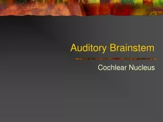
INFERIOR COLLICULUS Level • Inferior colliculus is a large nucleus of gray matter that lies beneath a corresponding surface elevation. • It is part of the auditory pathway. • It receives fibers from the lateral lemniscus. • Its efferent fibers pass to the thalamus
INFERIOR COLLICULUS Level • Trochlear nucleus: lies in the central gray matter close to the median plane just posterior to the medial longitudinal bundle. The fibers of the trochlear nerve decussate in the superior medullary velum. 2. Decussationof the superior cerebellar peduncles in the mid line.
INFERIOR COLLICULUS Level 3. Substantianigra: • Occupies the most ventral part of the tegmentum. • It consists of pigmented, melanin containing neurones. • It projects to the basal ganglia. Its degeneration is associated with Parkinson’s disease. Mask Face Flexion of the Trunk Pill-Rolling Tremors Slow Shuffling Feet movement
ASCENDING LEMINISCI • Composed Of: • Spinal (Lateral & anterior spinothalamic tracts) • Trigeminal (Lateral & medial). • Lateral lemniscus. • Medial lemniscus. • Position: • Deeply placed lateral to the medial longitudinal fasciculus.
CRUS CEREBRI • It is a massive mass ventral to the substantianigra. • It consists entirely of descending cortical efferent fibers (Frontopontine, Corticospinal & corticobulbar and TemporopontineFibres)to the motor cranial nerve nuclei and to anterior horn cells. • Involved in the coordination of movement.
SUPERIOR COLLICULUS Level • A large nucleus of gray matter that lies beneath corresponding elevation. • It forms part of the visual reflexes. • Its efferent fibers go to the anterior horn cells & to cranial nuclei 3, 4, 6, 7 & 11). • It is responsible for the reflex movements of the eyes, head and neck in response to visual stimuli, as in following a moving object or altering the direction of the gaze.
SUPERIOR COLLICULUS Level 1. Occulomotor nucleus: • Situated in the central gray matter close to the median plane. • The fibers of the occulomotor nerve passes anteriorly through the red nucleus to emerge on the medial side of the crus cerebri.
SUPERIOR COLLICULUS Level 2. Red nucleus : • A rounded mass of gray matter that lies between the substantianigra and the cerebral aqueduct in the central portion of the tegmentum. • Its red coloration is due to its vascularity and the presence of an iron containing pigment in the cytoplasm of its neurons. • It is involved in motor control.
RETICULAR FORMATION • It is a complex matrix of nerve fibers & small groups of nerve cells that extends throughout the brain stem and projects to thalamic nuclei that influence large areas of the cerebral cortex.
RETICULAR FORMATION • It has a number of important functions i.e. Respiratory and Cardio- vascular centers are located in the medullary and caudal pontine reticular formation. • Some reticular neurons have long ascending and descending axons that allow profuse interaction with other neuronal systems.
RETICULAR TRACTS • Reticulo spinal tracts: • Influence a muscle tone & posture • Reticular Activating system: RAS Midbrain portion of RAS most likely is its center • Formed of some of the ascending fibers of the reticular formation. • They activate the cerebral cortex through the thalamus.
RAS • Functions as a net or filter for sensory input. • Filters out repetitive stimuli. • Allows passage of infrequent or important stimuli to reach the cerebral cortex. • Unless inhibited by other brain regions, it activates the cerebral cortex – keeping it alert and awake.
RETICULAR NEURONES • RapheNuclei: • Midline reticular nuclei. • Its ascendingfibers to the cerebral cortex are involved in the mechanisms of sleep. • Its descending fibers to the spinal cord are involved in the modulation of Pain. • Locus Coeruleus: • Pigmented neurons that lie in the tegmentum of the caudal mid brain & rostral pons • It is the main noradrenergic cell group of the brain. The locus coeruleus is the principal site for brain synthesis of norepinephrine
Internal Features Midbrain at level of inferior colliculus shows the following: • Corticospinal fibers • Frontopontine fibers • Temporopontine fibers • Substantianigra • Decussation of superior cerebellar peduncle • Medial longitudinal fasciculus • Medial lemniscus • Spinal lemniscus • Lateral lemniscus • Nucleus of trochlear nerve • Trigeminal lemniscus
Internal Features Midbrain at level of superior colliculus shows the following: • Corticospinal fibers • Frontopontine fibers • Temporopontine fibers • Substantianigra • Red nucleus • Decussation of rubrospinal tracts • Medial longitudinal fasciculus • Medial lemniscus • Spinal lemniscus • Trigeminal lemniscus • Nucleus of oculomotor nerve
Clinical Notes Raised intracranial pressure and Arnold –Chiari malformation leads to: • Herniation of medulla and tonsils of cerebellum • Traction of the lower 4 cranial nerves • Paralysis of the above mentioned nerves
Lateral Medullary Syndrome (Wallenberg) • Occlusion of posterior inferior cerebellar artery • All structures supplied by this artery will be affected: 1-Nucleus ambiguous 2-Nucleus of spinal tract of trigeminal N 3-Vestibular nuclei 4-Descending sympathetic fibers 5-Inferior cerebellar peduncle
Medial Medullary Syndrome • Occlusion of medullary branch of vertebral artery • All structures supplied by this artery will be affected 1-Pyramidal tract 2-Medial leminiscus 3-Hypoglossal nerve
Pontine Hemorrhage • Pons is supplied by basilar, anterior inferior and superior cerebellar arteries • Unilateral occlusion of one of the above vessels will affect the 1-Facial nerve 2-Abducent nerve 3-Vestibular nuclei 4-Corticospinal tract 5-Trigeminal nerve nuclei 6-Cochlear nuclei 7-Medial and spinal leminisci 8-Middle cerebellar peduncle
Vascular Lesions in Midbrain • Weber’s syndrome • Occlusion of a branch of posterior cerebral artery • Affects oculomotornerve and crus cerebri • Benedikt’s syndrome • Affects red nucleus and medial leminiscus
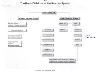
SUMMARY • The brain stem is composed (from above downwards) of: midbrain, pons & medulla oblongata which are continuous with each other, with diencephalon above & with spinal cord below. • The brain stem is connected with cerebellum through three pair of cerebellar peduncles. • The brain stem is the site of cranial nuclei, the pathway of important ascending & descending tracts & the site of emergence of cranial nerves (from 3rd to 12th). • Cranial nerves (with the exception of 4th) emerge from ventral surface of brain stem.