Download
1 / 26
300 likes | 544 Views
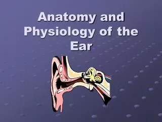
Anatomy of the Ear. Color Code Important Doctors Notes Notes/Extra explanation. Please view our Editing File before studying this lecture to check for any changes. Objectives. By the end of the lecture the student should be able to:

E N D
Anatomy of the Ear Color Code Important Doctors Notes Notes/Extra explanation Please view our Editing File before studying this lecture to check for any changes.
Objectives By the end of the lecture the student should be able to: • List the parts of the ear: External, Middle (tympanic cavity) and Internal (labyrinth). • Describe the parts of the external ear: auricle and external auditory meatus. • Identify the boundaries of the middle ear : roof, floor and four walls (anterior, posterior, medial and lateral). • Define the contents of the tympanic cavity: I. Ear ossicles,: (malleus, incus and stapes) II. Muscles, (tensor tympani and stapedius). III. Nerves (branches of facial and glossopharyngeal). • List the parts of the inner ear, bony part filled with perilymph (Cochlea, vestibule and semicircular canals), in which is suspended the membranous part that filled with endolymph). • List the organs of hearing and equilibrium.
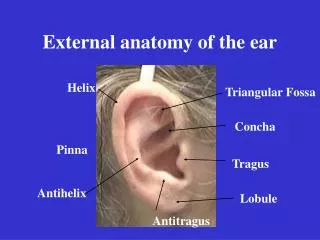
External Ear 05:18 • It is formed of the auricle, & the external auditory meatus. • The Auricle has a characteristic shape and collects air vibrations reception of sound. • It consists of a thin plate of elastic cartilage covered by a double layer of skin. • It receives the insertion of extrinsic muscles*, which are supplied by the facial nerve. • Sensation is carried by great auricular (from cervical plexus) & auriculotemporal (from mandibular) nerves. *these muscles are insignificant in humans because they don’t move but are prominent in animals, example: bunnies Extra
External Ear • The external auditory canal is a curved S-shaped tube about 2.5cm (one inch), that conducts & collects sound waves from the auricle to the tympanic membrane. Its outer 1/3rd is elastic cartilage, while its inner 2/3rds are bony. • It is lined by skin, and its outer 1/3rd is provided with hairs, sebaceousand Ceruminous Glands: (modified sweat glands that secrete a yellowish brownish substance called the ear wax). يمنع دخول الأتربة والحشرات الصغيرة Extra
Middle Ear (Tympanic Cavity) • Middle ear is a narrow, oblique, slit- like cavity (air-filled) in the petrous temporal bone & lined with mucous membrane. • It contains the auditory ossicles(the ear bones), which transmit the vibrations of the tympanic membrane (eardrum) to the internal ear. Extra
**you have to know all 3 names Middle Ear (Tympanic Cavity) • Communicates anteriorly with the Nasopharynx* through the Auditory Tube (also called pharyngotympanic or eustachian tube)**, which extends from the anterior wall downward, forward, and medially to the nasopharynx). • The posterior 1/3rd of the canal is bony, and its anterior 2/3rds are cartilaginous. (the external ear was the opposite) • Its function is to equalize the pressure on both sides of the ear drum. (normally it is closed but it opens to balance the pressure) *this is significant clinically because recurrent throat infections can travel to the ear Extra
Middle Ear (Tympanic Cavity) • The middle ear has: • Roof, • Floor, • and 4 walls: • Anterior, • Posterior, • Lateral, and • Medial. ROOF Posterior Anterior MEDIAL FLOOR
Middle Ear (Tympanic Cavity) Roof & Floor Extra • The Roof is formed by a thin plate of bone, called tegmen tympani, which is part of the petrous temporal bone. • It separates the tympanic cavity from the temporal lobe of the brain. The Floor is formed by a thin plate of bone, which separates the middle ear from the bulb of the internal jugular vein.
Middle Ear (Tympanic Cavity) Anterior Wall • The anteriorwall is formed below by a thin plate of bone that separates tympanic cavity from the internal carotid artery. • There are 2 canals at the upper part of the anterior wall. • The upper, smaller is the canal for the tensor tympani muscle. • The lower, larger is for the auditory tube.
Middle Ear (Tympanic Cavity) Posterior Wall • The posterior wall has in its • Upper part a large, irregular opening, theaditus to the mastoid antrum (a cavity behind the middle ear, within mastoid process, it contains air cells) • Below: a small, hollow, conical projection, the pyramid, which houses the stapedius muscle and its tendon. (The tendon emerges from the apex of the pyramid.) Extra
Middle Ear (Tympanic Cavity) Medial Wall • Greater part of the medial wall shows a rounded projection, (Promontory) that results from the underlying 1st turn of the cochlea. • Above and behind the promontory lies the Oval window*(Fenestra Vestibuli), which is closed by the base of the stapes. • Below and behind the promontory lies the Round window (Fenestra Cochleae). Which is closed by thesecondary tympanic membrane • It is formed by the lateral wall of the inner ear. *also called foramen ovale
Middle Ear (Tympanic Cavity) Lateral Wall • The lateral wall is largely formed by the tympanic membrane (its like a satellite to collect sound). • The membrane is obliquely placed, facing downward, forward, & laterally. • It is extremely sensitive to pain. • Nerve supply of ear drum: • Outer surface: • 1- Auriculotemporal nerve. • 2- Auricular branch of vagus. • Inner surface: • Tympanic branch of the glossopharyngeal nerve. Extra The lateral wall is toward the external ear The medial wall is toward the inner ear Extra
Middle Ear (Tympanic Cavity) Tympanic Membrane • Normally, It is concave laterally, and at the depth of its concavity there is a small depression, “ the Umbo” produced by the tip of the handle of the malleus. • When the membrane is illuminated through an otoscope*, the concavity produces a “Cone of Light," which radiates anteriorly and inferiorly from the umbo. • Most of the of the membrane is tense and is called the Pars Tensa. • Asmall triangular area on its upper part is slack and called the Pars Flaccida. * Pars Tensa tense end Pars Flaccida flaccid which means loose Extra Extra
Middle Ear (Tympanic Cavity) Auditory Ossicles • The auditory ossicles are 3: • Malleus (hammer), • Incus (anvil), • Stapes (stirrup). • They transmit sound waves from tympanicmembrane to the perilymph of the internal ear. • They are covered by mucous membrane & articulate by synovial joints*. *المشاكل في هذه المفاصل لدى كبار السن هي سبب ضعف السمع لدى بعضهم Extra
Middle Ear (Tympanic Cavity) Muscles of the Ossicles • Origin:Cartilage of the auditory tube and the bony walls of its own canal. • Insertion:into the handle of the malleus. • Nerve supply: Mandibular nerve. • Action: Contracts reflexly in response to loud sounds to limit the excursion of the tympanic membrane. TENSOR TYMPANI STAPEDIUS (the smallest voluntary muscle) • Origin: Internal walls of the hollow pyramid. • Insertion: The tendon emerges from the apex of the pyramid and is inserted into the neck of the stapes. • Nerve supply: Facial nerve. • Action: Reflexly damps down the vibrations of the stapes by pulling on the neck of that bone.
Middle Ear (Tympanic Cavity) Nerves • Tympanic nerve • It is a branch of the glossopharyngeal nerve. • It gives: • Tympanic plexus on the promontory • The tympanic plexus gives the, Lesser petrosal* nerve which relays in the otic ganglion. • It gives secretomotor supply to the parotid gland *Compare: Extra Extra
Middle Ear (Tympanic Cavity) Nerves • Facial nerve (VII) • Enters through the Internal acoustic meatus with the 8thvestibulocochlear nerve. • It expands to form Geniculate ganglion. • It passes vertical behind the pyramid. • It leaves the middle ear through the stylomastoid foramen. • Branches: • 1. Greater Petrosal nerve. • Arises from Geniculate Ganglion. • Carries preganglionic parasympathetic to supply: Lacrimal, Nasal, and Palatine glands. • 2. Nerve to Stapedius. • 3. Chorda Tympani: • Arises just before the facial nerve exits. Recall: chorda tympani carries taste fibers. So if there was any damage to this nerve the patient will experience dyspepsia Ex: during ear surgeries To remember: chorda chocolate ordates
Internal Ear, Or Labyrinth • Labyrinth is situated in the petrous part of the temporal bone, medial to the middle ear. • It consists of : • Bony labyrinth: • A series of bony chambers lined by endosteum. • They contain a clear fluid, the perilymph, in which is suspended the membranous labyrinth. • Membranous labyrinth: • consists of a series of membranous sacs and ducts within the bony labyrinth, it is filled with endolymph. Note: The middle ear was filled with air, but the inner ear is filled with fluid. In the bony labyrinth that fluid is perilymph and in the membranous labyrinth is it endolymph. Extra
Internal Ear (Labyrinth) Bony Labyrinth • The bony labyrinth consists of: • Cochlea • Vestibule, • Semicircular canals, • Cochlea • Its first turn produces the promontory on the medial wall of the tympanic cavity. • It contains the cochlearduct (part of the membranous labyrinth). Extra
Internal Ear (Labyrinth) Bony Labyrinth • Vestibule • Isthe central part of the bony labyrinth. • Contains the utricle & saccule (parts of the membranous labyrinth) • In the lateral wall of the vestibule are: • the fenestra vestibuli (oval window), which is closed bythe base of the stapes, and • the fenestra cochleae (round window), which is closed by the secondary tympanic membrane. Extra To remember: oval vestibuli round cochleae
Internal Ear (Labyrinth) Bony Labyrinth • Semicircular Canals • Semicircular canals: superior (anterior), posterior & lateral. • Each canal has a swelling at one end called the ampulla. • The canals open into the vestibule by five orifices, one of which is common to two of the canals. • Lodged within the canals are the semicircular ducts.
Internal Ear (Labyrinth) Membranous Labyrinth • The membranous labyrinth consists of (Four ducts & Two sacs) which are freely communicate with one another : • Sacs: Utricle & Saccule lodged in the bony vestibule. • Ducts: Three semicircular ducts lie within the bony semicircular canals. (anterior, posterior, lateral) • Cochlear Duct: lies within the bony cochlea. • The cochlear duct divides the bony cavity into • Scala Vestibuli (the perilymph is separated from the middle ear by the base of the stapes at the fenestra vestibuli) • Scala Tympani (the perilymph is separated from the middle ear by the secondary tympanic membrane at the fenestra cochleae) Only on the boys’ slides
Internal Ear (Labyrinth) Membranous Labyrinth • Located on the walls of the utricle and saccule are specialized sensory receptors, which are sensitive to the orientation of the head to gravity or other acceleration forces. • The utricle, saccule and semicircularducts are concerned with maintenance of Equilibrium. • The highly specialized epithelium on the floor of cochlearduct forms the Spiral organ of Cortithat contains the sensory receptors for Hearing.
MCQs 1. The outer 1/3rd of the external auditory canal is: A- bony B- elastic cartilage C- fibrous cartilage D- hyaline cartilage Answer: B 2. The auditory ossicles are found in: A- external ear B- middle ear C- internal ear D- labyrinth Answer: B 3. The tympanic cavity communicates with the nasopharynx via: A- laryngotympanic duct B- lacrimal duct C- internal acoustic meatus D- eustachian tube Answer: D 4. The floor of the middle ear separates it from the bulb of: A- internal jugular vein B- external jugular vein C- internal carotid aretery Answer: A 5. The auditory ossicles articulate by _____ joints: A- fibrous. B- cartilaginous. C- synovial. Answer: C 6. Stapedius is inserted into: A- handle of the malleus B- handle of the stapes C- neck of the stapes D- neck of the malleus Answer: C 7. Utricle & saccule are lodged within the: A- cochlea B- vestibule C- semicircular canal D- tympanic cavity Answer: B 8. Which of the following is responsible for hearing: A- utricle B- saccule C- semicircular duct D- cochlear duct Answer: D
Members: Hamad Alkhudairy Abdulrahmanalrajhi Leaders: NawafAlKhudairy Jawaher Abanumy Feedback anatomyteam436@gmail.com @anatomy436 References: 1- Girls’ & Boys’ Slides 2- Greys Anatomy for Students 3- TeachMeAnatomy.com Anatomy Team