Download
1 / 27
270 likes | 451 Views
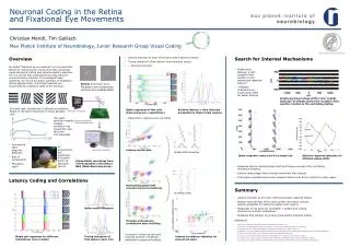
How the eye sees Last time Anatomy of the eye Cells in the retina Rods and cones Visual receptors This time Visual receptors Visual transduction. 1. Different cells in the retina. The Basic Retinal Circuit. Back of eye. 6. Pigment cells. 1. Receptor Cells (rods and cones)

E N D
How the eye sees Last time Anatomy of the eye Cells in the retina Rods and cones Visual receptors This time Visual receptors Visual transduction 1
Different cells in the retina The Basic Retinal Circuit Back of eye 6. Pigment cells 1. Receptor Cells (rods and cones) 2. Bipolar Cells 3. Ganglion Cells 4. Horozontal Cells 5. Amacrine Cells Structure of the eye Front of eye 2
Photoreceptor cells are the light sensors Back of eye Front of eye 3
The visual receptors are G Protein Coupled Receptors • seven transmembrane regions • hydrophobic/ hydrophilic domains • conserved motifs • chromophore stably attached to receptor (Schiff’s base Lys296 in TM7) • thermostable 4
Different opsins recognize different wavelengths We have 4 different opsins Rods: Rhodopsin: blue/green sensitive pigment Cones: S opsin: blue sensitive M opsin: green sensitive L opsin: red sensitive 5
The light catcher is 11-cis-retinal • covalently attached to opsin GPCR • Vitamin A derivative • Binds light, changes conformation from 11-cis to all-trans 6
Rhodopsins are packed in a crystalline array in the disc 8 10 rhodopsins/cell Atomic force microscopy 7
They even make other cells do their work: Pigment cells recycle retinal Photoreceptor Interphotoreceptor binding protein Carries retinal to pigment cell + Retinal modified to 11-cis Combines with opsin to form rhodopsin Pigment cell 8
Retinal Ganglion cells express melanopsin, are sensitive to light and project to the superchiasmatic nucleus 11
The visual cascade is a G protein-coupled cascade Rhodopsin Gtransducin phosophodiesterase cGMP to GMP close cGMP channels 16
High amplification in the visual cascade Rhodopsin Gtransducin phosophodiesterase cGMP to GMP close cGMP channels 1 100 100 100,000 1000? 19
Phototransduction is a highly regulated cascade Adapt to respond over 6 log orders of light 1. Long-term adaptation -pupil size -receptor photobleaching 2. Short-term adaptation -recovery of membrane potential -deactivation of receptors 20
Negative regulation of phototransduction Rhodopsin Gtransducin phosophodiesterase cGMP to GMP close cGMP channels Rhod kinase GAP Guanylate cyclase GTP to cGMP open channels Arrrestin Drop in Ca influx activates Ca dissociates from Calmodulin, Opens channels 21
Mice without GAP cannot turn off light response quickly no GAP with GAP (wild-type) 25
Phototransduction: Differences between rods and cones RodsCones Very sensitive to light 30x less sensitive to light each rhodopsin activates 30x less G proteins 26
Properties of phototransduction • responds to 1 photon of light • responds over a range of 6 log orders of light • responses are extremely reliable • 1000s of discs maximize surface area of light detection • high concentration and thermostability of rhodopsin means high detection, low noise • adaptation increases the operating range Photoreceptors are highly specialized to detect light! 27