Download
1 / 22
240 likes | 366 Views
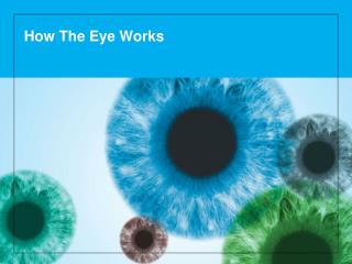
How The Eye Works. Insert name/ Practice name/ Logo here if desired. The healthy eye. Light rays enter the eye through the clear cornea, pupil and lens. These light rays are focused directly onto the retina, the light-sensitive tissue lining the back of the eye.

E N D
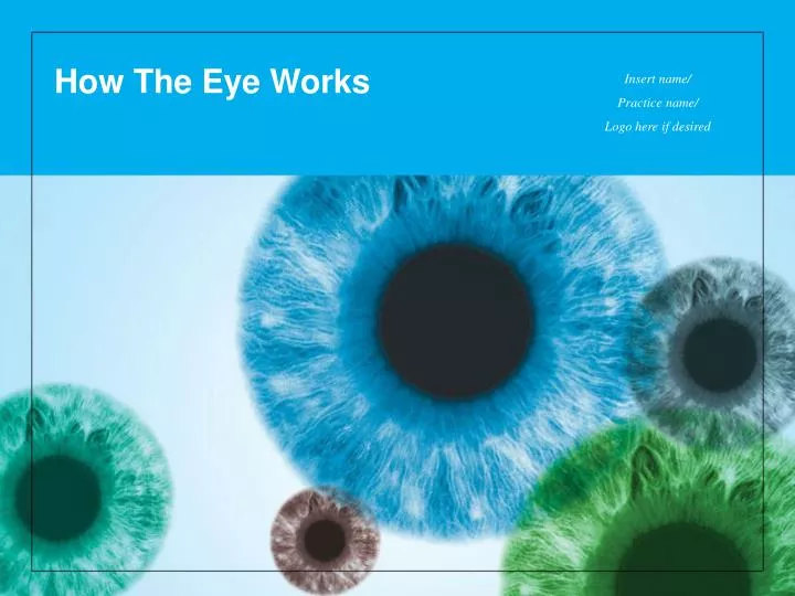
How The Eye Works Insert name/ Practice name/ Logo here if desired
The healthy eye • Light rays enter the eye through the clear cornea, pupil and lens. • These light rays are focused directly onto the retina, the light-sensitive tissue lining the back of the eye. • The retina converts light rays into impulses, sent through the optic nerve to your brain, where they are recognized as images. • 70% of the eye's focusing power comes from the cornea and 30% from the lens.
Refractive errors • Inability to see clearly is often caused by refractive error. • Four types of refractive error: • Myopia (nearsightedness) • Hyperopia (farsightedness) • Astigmatism • Presbyopia
Refractive errors: myopia • In myopia (nearsightedness), there is too much optical power in the eye; the distance between the cornea and the retina may be too long. • The distance between the cornea and the retina may be too long or the power of the cornea and the lens may be too strong. Myopia
Refractive errors: myopia • Close objects will look clear, but distant objects will appear blurred (hence, “nearsightedness”). Myopia, or nearsightedness
Refractive errors: hyperopia • In hyperopia (farsightedness), there is too little optical power. • The distance between the cornea and the retina may be too short. • Light rays are focused behind the retina instead of on it. Hyperopia
Refractive errors: hyperopia • In adults (but not necessarily children) distant objects will look clear, but close objects will appear blurred (hence, “farsightedness”). Hyperopia, or farsightedness
Refractive errors: astigmatism • In astigmatism, the cornea is curved unevenly—shaped more like a football than a basketball. • Light passing through the uneven cornea is focused in two or more locations. • Distant and close objects may appear blurry. Astigmatism occurs when light passes through uneven cornea
Refractive errors: presbyopia • Presbyopia is a normal condition in which your eyes gradually lose the ability to focus things up close. • When we are young, the lens in our eyes is flexible and is able to change focus easily between near and far objects, like an autofocus on a camera. • At around age 40, this flexibility naturally begins to gradually decrease, making it more difficult to see objects up close.
Correcting refractive errors • Eyeglasses are the most common methods of correcting refractive errors; they refocus light rays directly on the retina.
Correcting refractive errors • Contact lenses: Acting like eyeglasses, contact lenses float on the tear film that coats the cornea—they refocus light rays on the retina.
Correcting refractive errors • Refractive surgery: A variety of surgical procedures that permanently alter the eye such that light rays are refocused on the retina to improve vision. • The most common refractive surgical procedures are: • Laser In Situ Keratomileusis (LASIK) • Epithelial Laser In Situ Keratomileusis (Epi-LASIK) • Photorefractive Keratectomy (PRK) • Laser Epithelial Keratomileusis (LASEK) • Conductive Keratoplasty (CK) • Astigmatic Keratotomy (AK) • Radial Keratotomy (RK) • Phakic Intraocular Lenses (IOLs) • Accommodative IOLs and multifocal IOLs • Refractive Lens Exchange (RLE) • Intrastromal Corneal Ring Segments (INTACS)
Common eye disease • In addition to refractive errors, many kinds of eye disease can affect your sight; vision changes are not always evident right away.
Common eye disease Age-related macular degeneration (AMD) • May have many contributing factors, including the natural aging process; the macula may degenerate or atrophy over time, possibly affecting your central vision. • Early stage AMD may be hardly noticeable; usually a bilateral disease with one eye more affected than the other. • Symptoms: blurry vision; dark or empty areas in central vision; straight lines may look wavy. • No specific treatment for “dry” form of AMD; laser, photodynamic therapy (PDT), or anti-VEGF drugs may treat the “wet” form of AMD. With AMD, dark areas may appear in your central vision
Common eye disease Glaucoma • A leading cause of loss of vision and blindness in the United States, especially for older people. • Disease of the optic nerve; commonly due to increased intraocular pressure (IOP). When optic nerve fibers are damaged, blind spots develop. • Blind spots or loss of vision usually go undetected until the optic nerve is significantly damaged. • Treatment (all aimed at lowering intraocular pressure): eyedrops, laser surgery or conventional surgery to shunt fluid from the eye may be required, depending on the type of glaucoma and its stage. • Early detection and treatment are keys to preventing vision loss from glaucoma. Normal vision Vision as it might be affected by glaucoma
Common eye disease Diabetic retinopathy • Diabetes Mellitus is the inability of the body to use and store sugar properly, resulting in high blood sugar levels. • Results in changes in veins, arteries and capillaries in the body, including the eyes. Damage occurs to the fragile blood vessels of the retina. • Symptoms: blurred, decreased vision, loss of vision. With diabetic retinopathy, cholesterol or other fat deposits from blood, called hard exudates, may leak into the retina
Common eye disease Diabetic retinopathy • Treatment: Usually laser surgery; occasionally conventional surgery. • You can significantly lower your risk of vision loss by maintaining strict control of your blood sugar level and frequent visits to your ophthalmologist.
Common eye disease Cataract • Age-related cataract is the most common form. • The eye’s normally clear lens becomes cloudy, preventing light from focusing sharply on the retina. • Symptoms: blurry vision; glare or light sensitivity; poor night vision; difficulty driving at night; yellowing or fading of colors; increased light required to read comfortably. Yellowing of colors is a symptom of cataract
Common eye disease Cataract • Treatment: surgery removes the cloudy lens and replaces it with an artificial intraocular lens implant (IOL). • If cataract symptoms are not adversely affecting your daily activities, you may not need surgery. (Simply have eyeglass prescription changed as needed.)
Preserve good vision with regular visits to an ophthalmologist or other medical professional • Infants and young children should visit an ophthalmologist or other medical professional at the following intervals: • Newborn to 3 months • 3 to 6 months • 6 months to one year • 3 to 3 1/2 years • 5 years • Other medical professionals include pediatricians, family physicians, nurse practitioners or physician assistants.
Preserve good vision with regular visits to an ophthalmologist or other medical professional • Age-appropriate eye and vision evaluations should be performed in the newborn period and at all subsequent health supervision visits, since different childhood eye problems may be detected at each. • The screening process includes a history in order to assess risk factors as well as an examination. • Vision testing should be performed for a child at the earliest age that is practical, and it is recommended for all children starting at 3.
Preserve good vision with regular visits to an ophthalmologist Visit your ophthalmologist at the following intervals: • Age 20-29 years: At least once during this period • Those with risk factors for glaucoma (people of African descent or those who have a family history of glaucoma) should be seen every 3-5 years. • Age 30-39 years: At least twice during this period • Those with risk factors for glaucoma (people of African descent or those who have a family history of glaucoma) should be seen every 2-4 years. • Age 40-64 years: Every 2-4 years. • Age 65 years or older: Every 1-2 years. • People with diabetes should visit their ophthalmologist once a year.