Download
1 / 4
40 likes | 42 Views
You need to get in touch with the best retina specialist in Mumbai if you are facing any kinds of eye problems to detect if there is a problem in any part of the retina, so as to treat it at the earliest.<br><br>Read more @ http://bit.ly/2SmWySr

E N D
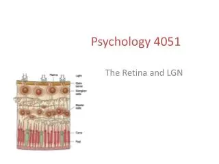
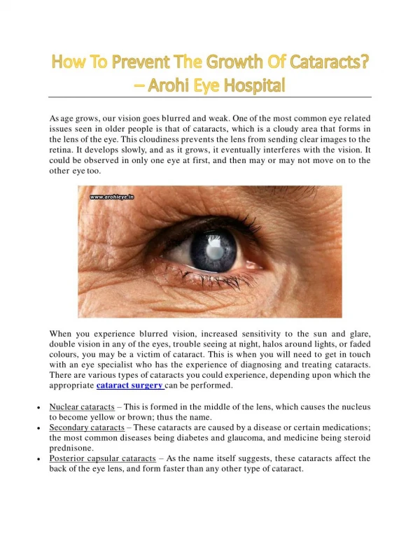
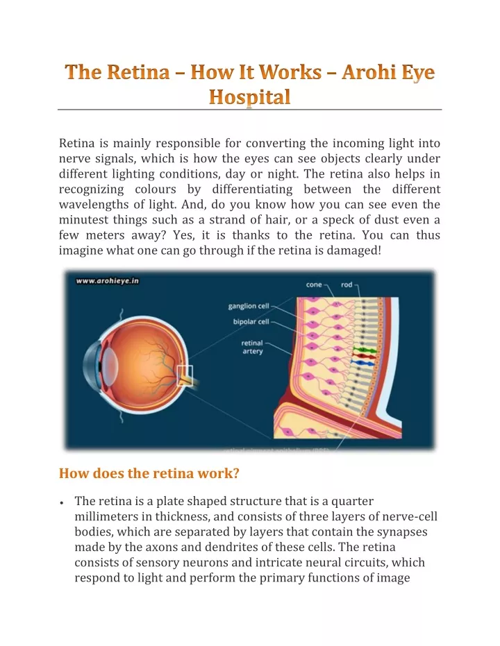
Retina is mainly responsible for converting the incoming light into nerve signals, which is how the eyes can see objects clearly under different lighting conditions, day or night. The retina also helps in recognizing colours by differentiating between the different wavelengths of light. And, do you know how you can see even the minutest things such as a strand of hair, or a speck of dust even a few meters away? Yes, it is thanks to the retina. You can thus imagine what one can go through if the retina is damaged! How does the retina work? The retina is a plate shaped structure that is a quarter millimeters in thickness, and consists of three layers of nerve-cell bodies, which are separated by layers that contain the synapses made by the axons and dendrites of these cells. The retina consists of sensory neurons and intricate neural circuits, which respond to light and perform the primary functions of image
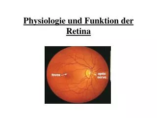
processing respectively. Then, an electrical message travels through the optic nerve to the brain, which further processes the visual perception. The retina has a number of photoreceptors at the back, which contain pigment molecules that are excited when light touches them. These photoreceptors are in touch with the epithelial layer of the eye, which provide a steady stream of retinal molecules. These molecules, when exposed to light, undergo a conformational change, and are recycled back into the pigment epithelium, which contain melanin granules that absorb stray photons, preventing them from creating a reflection on the photoreceptors, which makes the images appear blurred. There are two types of photoreceptors – the rods used for low light vision, and the cones for daylight and bright coloured vision. Can you imagine that such a small human retina consists of 4-5 million cones, and 77-107 million rods? And, the brightest vision can be experienced when the light directly falls on the Macula region of the retina, which consists of a large number of small and tightly packed cones. Each photoreceptor cell consists of an outer photopigment segment, inner mitochondria segment, a nucleus, an inner fiber, and a synaptic terminal. The photoreceptor cell is connected to the ganglion cell layer through bipolar cells; and the rods and cones provide input to these bipolar cells. These ganglion cells transmit the visual information from the retina to the brain. The most common retinal diseases There are various kinds of diseases and disorders that can harm the retina, which affect the vision, making it blurred or distorted. The most common retinal diseases include –
Retinal tear or detachment – This damage is caused when the vitreous moves away from the retina so much that it tears the retina. Fluid tends to pass through this retinal tear, thus lifting it off the back of the eye. With such a vision becomes blurred, and may also result in blindness. retinal detachment , the Macular hole – This condition is caused due to the shrinkage or separation of the vitreous, causing sudden decrease in vision. The various reasons that can cause macular hole include diabetic eye disease, retinal detachment, eye injury, macular pucker, high amount of nearsightedness, or an inherited condition. Macular degeneration – Macular degeneration occurs when parts of the macula get thinner with age, causing tiny clumps of proteins to grow on them. This disorder results in losing the central vision, which does not let the patient identify the fine details of an object. But, the peripheral/side vision remains normal. Macular degeneration may also occur when there is abnormal growth of blood vessels under the retina, causing blood or other fluid to leak from these vessels, thus causing macula scarring. This is a rare but serious type of macular degeneration, which brings vision loss much faster. Diabetic retinopathy – This disorder affects those suffering , as the high blood sugar levels often damage the from diabetes blood vessels within the retina, causing them to swell and leak. And, if they completely close, blood flow is completely stopped! Retinal vein occlusion – This disorder is caused when the veins that carry blood away from the retina get blocked due to hardened arteries which cause blood clots. The various factors that can cause retinal vein occlusion include diabetes, high blood pressure, atherosclerosis, and other eye conditions like macular edema, glaucoma, and vitreous hemorrhage.
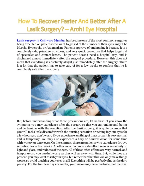
To stay away from all such disorders and damages, it is important that you have regular visits to an eye specialist. Arohi Eye is the Hospital best retina specialist in Mumbai can have your eyes examined by expert doctors to determine early symptoms of any of the above or other retinal diseases, so that the right actions can be taken before the condition gets worse. where you Follow us on: Facebook, Twitter, Instagram & YouTube