Download
1 / 11
110 likes | 129 Views
The Eye. A Model for Sensing. Three components: Stimulus detection – a specialized sensory neuron Reception – where neurons receive information from the sensory neurons Integration – where information from receivers is processed All the human sensory systems have these components.

E N D
A Model for Sensing • Three components: • Stimulus detection – a specialized sensory neuron • Reception – where neurons receive information from the sensory neurons • Integration – where information from receivers is processed • All the human sensory systems have these components.
The Visual System • Retina • Optic nerve (axons of ganglia in eye) • Layers in the thalamus (LGN) • Secondary paths to SCN (circadian rhythms) and superior colliculi to control eye movement • Primary visual cortex
Adaptation • Adaptation -- diminishing receptiveness of a sensory receptor neuron. • Habituation – also diminishing receptiveness but at a different level (within a neuronal circuit not a cell). • Adaptation is essential for the perception of change. • Dark and light adaptation permit vision under different environmental conditions.
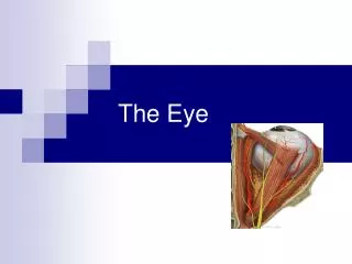
Parts of the Eye • Cornea – protects eye and initiates focusing • Lens – focuses light on the appropriate spot on the retina • Iris – adjusts focus by opening and closing pupil to admit light • Pupil – hole that admits light
More Parts of the Eye • Aqueous and vitreous humor – fluid inside eye • Sclera – whites of eye • Retina – layer of photoreceptors at the back of the eye, responsive to light • Blind spot – place where optic nerve exits the eye • Fovea – spot of best focus and densest cones
Types of Photoreceptors • Rods – used for brightness perception and motion • Cones – used for color and form (shape) perception • Cones are wavelength-specific: • Blue = 430 nm • Green = 530 nm • Red = 560 nm • Mixing all three equally produces white
Transduction • Photoreceptors release the neurotransmitter glutamate (glu) when depolarized. • Depolarized in the dark. • Hyperpolarized by light. • Only ganglion cells have action potentials. • Photoreceptors produce graded response that provides input aggregated by bipolar cells. • Magno ganglion cells receive input from rods, parvo ganglion cells from cones
Bipolar Cell Receptive Fields • The receptive field is the area of the retina capable of changing the bipolar cell’s membrane potential • Two kinds of receptive fields: • OFF cell – excitatory • ON cell – inhibitory • OFF and ON refers to light, not the cell • Center and surround are opposites
Edge Detection • The center-surround organization of the receptive fields of ganglion cells exaggerates the contrast at borders. • Visual processes “fill in” what occurs between borders (edges). • Contrast effects occur because we notice variations, not absolute magnitudes of light.
Color Contrast • Cones respond to specific wavelengths of light that determine hue. • Color cells have complementary surrounds that heighten contrast and strengthen their signal. • Opponents are: red/green, blue/yellow.