Download
1 / 33
330 likes | 448 Views
Corresponding Positions in SMOA Structure of Amino Acid Side Chains of known function in PHBH . SMOA and PHBH Residues 51-101. Positions of PHBH H72 SMOA Y73 (coil between strand-4 and helix-3) . SMOA Residues 101-136; PHBH Residues 101-152.

E N D
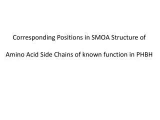
Corresponding Positions in SMOA Structure of Amino Acid Side Chains of known function in PHBH
SMOA and PHBH Residues 51-101 Positions of PHBH H72 SMOA Y73 (coil between strand-4 and helix-3)
SMOA Residues 101-136; PHBH Residues 101-152 Similar Positions of PHBH E104(Helix-6) SMOA L103(Helix-5)
SMOA Residues 101-136 and PHBH resudues 101-152 Overlap of Helix-5 of SMOA and Helix-6 of PHBH; Overlap of Strand-5 of SMOA and Strand-6 of PHBH; Superposition of PHBH antiparallel sheet (Strands 7,8,&9) with SMOA Helix-6 SMOA loop between Strand-6 and Helix-7 piercing adenosine ring. Possiblly related to low FAD-binding affinity. May also suggest an alternate FAD binding conformation than that observed in 1cc6.
SMOA residues 153-201; PHBH residues 136-215 PHBH Asp 286 (Helix-12) forms H-bond to OH ribose C-3. Similar position of Asp295 SMOA (Coil Following strand -14)
SMOA residues 153-201; PHBH residues 136-215 Similar positions of PHBH Y201(Strand-12) and SMOA M186(Strand-9) Strand -7 of SMOA shown in red introduces a gap in the primary sequence alignment
SMOA residues 201-248 and PHBH residues 215-247 Cool Substitution of SMOA A209 (Strand-10) For PHBH R220 (Strand-14) and SMOA V211(Strand-10) for PHBH Y222(Strand-14) in possible styrene-binding pocket
SMOA Residues 301-362; PHBH residues 291-351 Similar positions of PHBH P293(Loop between Helix-12 and Helix-13) and SMOA P302 (Loop between Strand-14 and Helix-13) Also similar positions of PHBH L299 and SMOA A308 in Helix-13
Corresponding structural Positions of fully conserved amino acids present in the Aligned Primary Sequencs of SMOA and PHBH
Close spatial relationship of conserved side chains in the first 50 amino acids
Conserved Amino acids in the sequence range 50-100 are close neighbors and associated with similar elements of secondary structure
Side Chains conserved in the primary sequence are in close proximity. Up until Ala 123, they also derive from similar secondary structural elements. Interestingly, the two pairs of leucines deriving from the positionally related helix of SMOA and three stranded antiparallel sheet of PHBH are still in close proximity and pointing in roughly the same direction.
The conserved side chains in the region 150-201 appear in non-overlapping positions in the SMOA and PHBH structures and derive from different elements of secondary structure in each protein. Interestingly, the alpha carbon chain-traces are still very similar in both structures.
Amino acids conserved in the primary sequence are distally located and derive from different secondary structural elements in this region of the three dimentional stucture
Very Similar Chain Trace; Conserved aa in primary sequence are still distally located in the 3-D structure in this region
Identical residues in the primary sequence alignment are once again in nearly identical positions. The proteins have returned from improvization mode in these helicies.
The C-terminal conserved residues of SMOA and PHBH are more divergent but positioned in similar secondary structure except for the two most C-terminal helicies of SMOA, which do not exist in PHBH
N-Terminal region of SMOA: Coil pierces through the flavin isoalloxazine ring. Suggests Flavin/protein may adopt an alternate configuration in the associated complex. This may contribute to the low affinity of oxidized FAD to SMOA.
Significant differences in the SMOA(Helix) and PHBH(Sheet) secondary structure. Also steric problem with SMOA loop piercing through adenosine ring. Suggests an alternate FAD-binding mode in SMOA. This may contibute to the weak binding of Oxidized FAD to SMOA.
C-Terminal Helicies are Unique to SMOA. May be a good place to add a tag or fuse SMOB.
Alternate FAD-binding configuration observed in 3C96 allows FAD to dock without piercing of the adenosine ring Alignment of SMOA and (3c96). Same central and C-terminal helix differences observed in comparing SMOA to PHBH
Docking in the FAD from the PHBH (1k0i) structure into the active site: Isoaloxazine ring of FAD is nolonger Pierced by SMOA. Adenosine Ring is still Pierced. No room for second PHB.
SMOA Compared with PHBH(1K0J) SMOB backbone structure does not pierce through the flavin ring system in this alignment. Also there is nearly room for NADPH to Fit in the SMOA active site
Alignment of SMOA and (2dki) Flavin docks with out steric conflict. Xenon atom in structure shown in green.
Superposition of (2qa2) and SMOA. Extra C-terminal domain red. Helix sheet mismatch orange. SMOA pierces isaloxazine ring of Docked from (2qa2) but does not clash with adenosine ring.
SMOA Superimposed with 2HD8. Flavin and styrene from 2HD8 docked into SMOA structure. Adeniosine and isalloxazine rings pierced by SMOA.
SMOA superimposed with (3c4a). Better homology for SMOA helix-6 than in any of the other related structures, which have three-stranded antiparallel sheets in this region. SMOA pierces docked isaloxazine ring but does not clash with adenosine ring.
Possible Sidechains lining the SMOA active site interacting with FAD and Styene