Download
1 / 95
980 likes | 1.06k Views
Metabolism of Amino Acid. SectionⅠ Physiological function and nutritional value of protein. Physiological functions of protein can not be replaced by carbohydrates or lipids 2. Nitrogen balance 3. Nutritional value of proteins: quantity + quality.

E N D
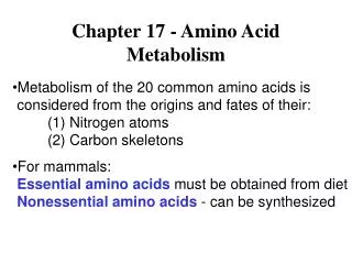
SectionⅠ Physiological function and nutritional value of protein • Physiological functions of protein • can not be replaced by carbohydrates or lipids • 2. Nitrogen balance • 3. Nutritional value of proteins: • quantity + quality
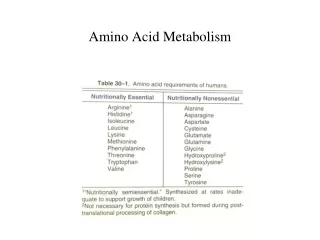
♣ Essential amino acids (8 kinds): * Can not be synthesized by human body * Must be supplied in the diet ♣ Non-essential amino acids(12 kinds) Nutritional value of proteins is related to the ratio of essential amino acids and non-essential amino acids
Made by complex routes (in bacteria & plants) Made by simple reactions
Section II Digestion, absorption and putrefaction of proteins
I. Digestion of DietaryProtein Self-activation of pepsinogen to active pepsin Trypsin Chymotrypsin Elastase Carboxypeptidase A、B pancreas Endopeptidase Exopeptidase Dipeptidase
II. Absorption of amino acid • Site: small intestine • Mechanism • 1. Na+-dependent carrier proteins: • Different carriers involve in absorption of different amino acids. • 2. γ-glutamyl cycle
γ-glutamyl transferase γ-glutamyl cycle
III. Putrefaction of protein The decomposition of the unabsorbed proteins, peptides or amino acids by anaerobic bacteria in the large intestine is termed putrefaction. • The resulting products are mostly toxic (amine, ammonia, phenol, indole and sulfureted hydrogen), most of which are excreted in feces.
co2 • (1) Formation of amines • Pr AAs amines (His,Lys,Trp,Tyr,Phe) • producing false neurotransmitters. • (2) Formation of ammonia • AAs NH3 + keto acid • blood urea NH3 • (3) Others • tryptophan → indoleand methylindole. • cysteine → H2S, Phenol etc. deaminase urease
Phe Tyr CO2 CO2 tyramine phenylethylamine (0H) (0H) Phenylethylatamine 苯乙醇胺 β- hydrotyramine β-羟酪胺(鱆胺)
Sources of amino acids Section Ⅲ General Metabolism of Amino Acids * Dietary proteins (the main source for our body) - digestion & absorption * Tissue proteins - degraded by proteolytic enzymes in lysosome - degraded by ubiquination in cytoplasm * Synthesis of non-essential AA in vivo
Ⅰ. Degradation of Tissue Proteins • Protein Turnover: • The process of continuous degradation and resynthesis of proteins. Proteins differ significantly in their turnover rates, which are measured in half-lives. • Significances: • Metabolic flexibility(灵活性): key regulatory enzymes, peptide hormones, receptor molecules; • Protects cells from the accumulation of abnormal proteins; • Numerous physiological processes are dependent on timely degradative and synthetic reactions: eukaryotic cell cycle control and antigen presentation.
Pathway of tissue proteins degradation • Protein degradation pathway in lysosomes • occurs inlysosomes • ATP independent • Non-specific protein degradationfor extracellular proteins membrane proteins long half-life intracellular proteins
Pathway of tissue proteins degradation • Ubiquitin—proteasome degradation pathway • occursin cytosol/nucleus • ATP dependent • Specific protein degradation for abnomal proteins short life proteins
Ubiquitination * Ubiquitin: 8.5KD small protein of 76 aas * 泛素活化酶(ubiquitin activating enzyme, E1) * 泛素结合酶(ubiquitin coupling enzyme,E2) * 泛素-蛋白连接酶(ubiquitin-protein ligase, E3) * 蛋白酶体(proteasome)- huge complex
ubiquitination Active Pr
Proteasome:huge complex • Proteasome exist in cell nuclear and cytoplasm, degrade abnormal and short-life proteins. 2α loop: 7 subunits/loop 2β loop: 7 subunits/loop 20S core particle (CP) 19S regulatory particles (RP):18 subunits,6 subunits have ATPase activity. 26s protein complex
Ⅱ. Amino acid metabolic pool Tissue protein Dietary protein Other pathways: Nitrogen-containing Compounds, one-carbon units, etc. Amino acid metabolic pool Tissue protein Synthesis of Nonessential amino acids Sources and fates of amino acids
excretion from urine ammonia urea in liver *deamination non-essential aa α-ketoacid CO2 +H2O + ATP Glc or lipids amine non-essential aa *decarboxylation CO2 Fates of amino acids
Ⅲ. Deamination of amino acids Amino acids R –C– COOH NH2 R –C– COOH O ammonia
The first step in the catabolism of AAs is removing of their α-amino group. Four types of deamination: 1. Transamination 2. L-Glu oxidative deamination 3. Associated deamination* -transamination associated with oxidative deamination -transamination associated with purine nucleotide cycle 4. Amino acid oxidase deamination
1. Transamination (Transaminase) Coenzyme: Pyridoxyl phosphate (PLP) (VitB6)
Distribute widely • Specificity of aminotransferases • Reversible reaction: • degradation of amino acids • amino acids synthesis • Can not remove free ammonia from molecular
The most important aminotransferases are to catalyze amino transfer between L-Glu and α-ketoacid:
Alanine aminotransferases (ALT) / glutamate-pyruvate transaminase (GPT)
Aspartate aminotransferase (AST) • / glutamate-oxaloacetate transaminase (GOT) COOH HC CH2 COOH NH2 α- ketoglutarate Asp AST COOH C=O CH2 COOH glutamate oxaloacetate
The activities of ALT and AST in different normal adult tissues
Diagnosis value of serum aminotransferases Two important enzymes in the clinical diagnosis of human disease are serum AST and ALT- abundant in heart and liver (intracellular enzymes). They are released as part of the cell injury that occurs in myocardial infarction, infectious hepatitis, or other damage to either organ. Assays of these enzyme activities in blood serum can be used both in diagnosis and in monitoring the progress of a patient during treatment.
Pyridoxal phosphate-derivedfrom vitamin B6, is the coenzyme of transaminase serving as transporter of amino group in the reaction. their coenzyme
The functional part of pyridoxal phosphate is an aldehyde functional group attached to a pyridine ring. The mechanism of transamination
2. L-Glu oxidative deamination L-Glutamate dehydrogenase ♣ Reversible • active in most tissue except skeletal and cardiac muscles; • allosteric enzyme (ATP/GTP inhibits, ADP/GDP activates).
3. Transamination associated with oxidative deamination (Associated deamination in most tissues) a-ketoglutarate NH3+NADH+H+ amino acid glutamate dehydrogenase Transaminase a-keto acid H2O+NAD+ glutamate Degradation of amino acids
NH3+NADH+H+ a-ketoglutarate amino acid glutamate dehydrogenase Transaminase a-keto acid H2O+NAD + glutamate Synthesis of nonessential amino acids
4. Transamination associated with purine nucleotide cycle in skeleton muscles and other tissues
5. Oxidative deamination of AAs ♣ (In kidney and liver cells)
deamination NH3 O R-CH-COOH α-ketoacid
Ⅱ. Fates of the carbon atoms of amino acids after deamination: • Be oxidized to produce CO2+H2O+ATP via citric acid cycle and oxidative phosphorylation. • Be converted into non-essential amino acids by amination. • Be converted into glucose, lipids or ketone bodies. glucogenic amino acids ketogenic amino acids ketogenic and glucogenic amino acids
Ketone bodies glucose
SectionⅣ Metabolism of Ammonia • Ammonia is extreme toxic compound to central nervous system. • The major way for detoxification of ammonia is to be converted into water-soluble, nontoxic urea in the liver, which is mainly passed via the bloodstream to the kidneys and excreted in the urine.
glutaminase Ⅰ. Sources of ammonia in the body: 1. Deamination of amino acids and amine. 2. Large quantity of ammonia is produced in intestine. (1) putrefaction of dietary protein (2) hydrolysis of urea: blood urea diffuse into intestine, degraded by bacterial urease. The ammonia is re-absorbed into the blood → liver. NH4+ (ammonium ion) < == > NH3 + H+ (ammonia) So alkaline soapsuds can NOT be used in clinical clyster for victim with hyperammonemia. 3. Reabsorbed from kidney in which ammonia is produced by hydrolysis of glutamine by glutaminase glutamine glutamate + NH3
cell Blood urine Gln Gln NH3 NH3 glutaminase Glu Glu Acidification of urine NH3 NH3 H+ NH3 NH4+
Ⅱ. Transportation of ammonia • The ammonia need to be • transported from the generated • tissue to the liver in a nontoxic • form. • 1. Alanine-Glucose cycle: • To transport ammonia in the nontoxic form of alanine from muscle to liver. • To regulate blood glucose indirectly and supply available glucose for muscle.
2. Transportation of ammonia with glutamine:Glutamine is a major transport form of ammonia. In brain or muscle COOH CH NH2 + NH3 (CH2) 2 COOH CONH2 CH NH2 (CH2) 2 COOH ADP+Pi Gln synthetase ATP glutaminase In liver or kidney Glu Gln In liver, NH3 urea In kidney, NH3 + H+ NH4+ is excreted in the urine.