Download
1 / 37
380 likes | 541 Views
The Structure & Function of Deoxyribose Nucleic Acid. Crash Course in The Why’s & How’s of DNA. Fredrick Griffith 1928. Frederick Griffith. Oswald Avery 1944. Hershey & Chase 1952. Hershey & Chase. The Race. Following Hershey & Chase, The 1950s became Obsessed with finding

E N D
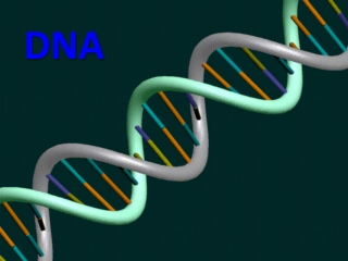
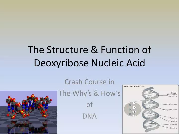
The Structure & Function of Deoxyribose Nucleic Acid Crash Course in The Why’s & How’s of DNA
The Race Following Hershey & Chase, The 1950s became Obsessed with finding DNA’s Structure! Finding the structure = Finding the function = Nobel Prize + Forever Notoriety
WATSON and CRICK • Announced in 1953 • Used the results of other scientists to figure out the structure of DNA
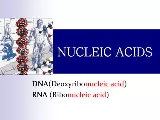
The Work of Biochemists Nitrogenous Base Phosphate Sugar
Franklin & Wilkins
Watson & Crick Model • Chemists found that DNA polymerized through the formation of phosphodiester linkages • This concluded a sugar-phosphate backbone • By analyzing the total number of purines and pyrimidines it was found that the number of A’s and T’s were equal to the number of C’s and G’s • This was called Chargaff’s rule after Erwin Chargaff • X-ray diffraction showed a repeating scatter pattern (.34 nm, 2.0nm, 3.4nm) • This repeating pattern only makes sense if the molecule is shaped as a double helix
Scatter Pattern X-ray Diffraction • Watson & Crick began to analyze the size and geometry of deoxyribose, phosphate groups, and nitrogenous bases. • Using things like bond angles, and measurements, they were able to devise 2.0nm probably represented the width of the helix, and .34 was likely the distance between bases stacked in the spiral • They arranged two strands of DNA running in opposite directions (5`-3` and 3`-5`)
And the Winner IS… Watson & Crick & Wilkins & NOT FRANKLIN
DNA Size • Width of the helix = 2.0nm • Length of one full complete turn of helix = 3.4nm • Distance between bases = .34nm • Antiparallel Double Helix • 3’ 5’ & 5’ 3’
The Roof of the Sugar Molecule C=O It is pointing at the 5’ end!
Base Pairing • Using the x-ray diffraction patterns and measurements, it was found only to work if: • Adenine always bonded with Thymine • Guanine always bonded with Cytosine • This phenomena is called Complimentary Base Pairing
DNA REPLICATION • Occurs during S-phase of the cell cycle • DNA has a special “complimentary structure that acts as a template for reproduction • This means, it allows for simple DNA copying • The strand unzips, and the old strand acts for a model to create a new “compliment” • The strand copies in two directions: • The Leading strand starts at the 3’ end and moves towards the 5’ end • The Lagging strand pieces together new nucleotides starting at the replication fork and works toward the 5’
Prokaryotic Replication Proteins • Helicase: Unwinds parental double helix at replication fork • Single Strand Binding Protein: Binds to and stabilizes DNA strand after it has separated • Topoisomerase: Relieves the “overwinding” strain that can occur ahead of the replication fork (swivel motion) • Primase: Synthesizes an RNA primer at the 5’ end of replicating strand. • DNA pol III: Using the template strand, covalently bonds nucleotides to the 3’ end of the pre-existing RNA strand or primer • DNA pol 1: Removes RNA primers & replaces them with DNA nucleotides • DNA Ligase: Joins 3’ end of DNA that replaces primer to the rest of strand (Joins Okazaki fragments of lagging strand)
DNA Excision Repair • Nucleotide excision repair (NER) is a particularly important excision mechanism that removes DNA damage induced by ultraviolet light (UV). • Recognition of the damage leads to removal of a short single-stranded DNA segment that contains the lesion (By the Nuclease enzyme). • The undamaged single-stranded DNA remains and DNA polymerase uses it as a template to synthesize a short complementary sequence. • Final ligation to complete NER and form a double stranded DNA is carried out by DNA ligase.
How Does a Cell Make Proteins? • The RNA molecule comes out of the nuclear envelope after it is transcribed from DNA (Its like a photocopy) • Transcription is the process of creating an RNA strand from a template of DNA nucleotides • The process of protein synthesis is called translation • Translation refers to the process of converting the “3-nucleotide RNA codons” into amino acids and then into amino acid chains
RNA & Protein Synthesis • RNA has very specific blue prints that it uses to build the various amino acids (proteins) called Codons. • These codons allow for the difficult job of the synthesis of the many proteins to be grouped into simple readable prints (codons) including “stop and start” codons
The Genetic Code • It is almost as if a cell is “pre-programmed” with a guide for making life • If there was such a “program” it would need to be something contained in nearly EVERY cell, so that each cell could individually work at it • We call this program “the Genetic Code” • It is the control for life as we know it