Download
1 / 31
410 likes | 849 Views
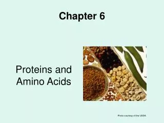
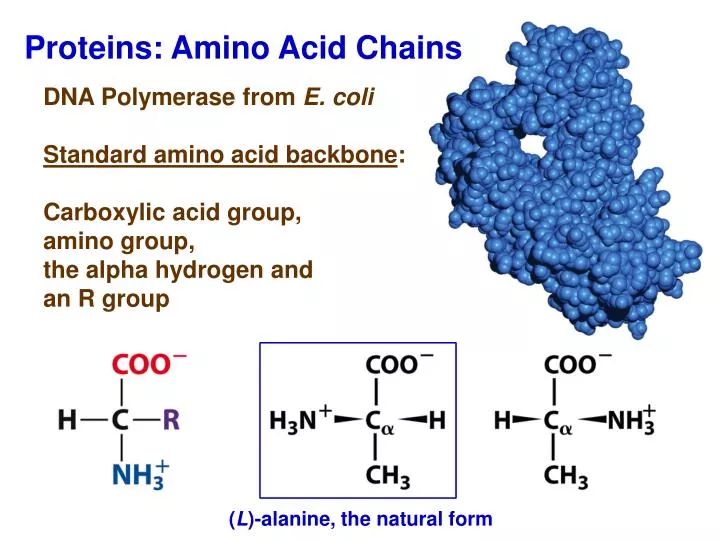
Proteins: Amino Acid Chains. DNA Polymerase from E. coli Standard amino acid backbone : Carboxylic acid group, amino group, the alpha hydrogen and an R group. ( L )-alanine, the natural form. Importance of Chirality in Biological Systems.

E N D
Proteins: Amino Acid Chains DNA Polymerase from E. coli Standard amino acid backbone: Carboxylic acid group, amino group, the alpha hydrogen and an R group (L)-alanine, the natural form
Importance of Chirality in Biological Systems (L)-thalidomide is an effective sedative for expectant mothers while (D)-thalidomide causes severe birth defects (L)-
Hydrophobic Amino Acids Non-polar side chains that interact very weakly with water. What type of bonding forces might contribute significantly for these AAs?
Polar Amino Acids Amino acid side chains readily interact with water Where might these AAs be located in a polypeptide?
Charged Amino Acids Always charged under physiological pH What is the predicted pKa values for these acids and bases?
Ionizible Amino Acids at Physiological pH Values Thiol group Cysteine (Cys) Thiolate anion Histidine (His) Imidazolium ion Are these oxidation/reduction reactions?
pKa Values of Ionizable Amino Acids ca. pKa 3 4 4 6 8 9 10 10 12
Disulfide Bond Formation Polypeptide stabilization Oxidation/reduction reaction
Amino Acid Coupling via Dehydration Synthesis What is the Nucleophile, Electrophile and Leaving Group in this reaction?
A polypeptide Primary Structure: Amino Acid Order or Sequence Coding convention: N- to C-terminus from left to right
Bond Length Indicates a Hybrid Bond Number of ca. 1.5 C-N single bond 1.45 Å C=N double bond 1.25 Å Peptide resonance
Peptide Bond Forms a Planar Unit Steric hindrance favors trans configuration
Ramachandran Diagram Shows Permitted Angles in Green +120 - 60 What is phi and psi?
α-Helix is a Coiled Polypeptide: Secondary Structure • What is the environmental condition favorable for a polypeptide to form an alpha helix? • Where in the polypeptide would an α-helix be located?
Beta-Sheet Polypeptide: Secondary Structure Antiparallel Parallel Which configuration would be more stable?
Beta-Sheet Backbone Is the distance of 7 Å reasonable? What do the green spheres represent? What is the green spheres orientation relative to the β-sheet? What is a favorable environment for beta-sheets formation?
Beta-Sheet Configurations: Super-Secondary Structure Beta-Barrel Reverse Turn Twisted-Sheet
CD4 Surface Protein in HIV with Four Similar beta-Sheet Domains
Alpha-Helix Configuration: Super-Secondary Structure • Common motif in DNA-binding proteins
Overall Configuration of a Single Polypeptide: Tertiary Structure Oxygen Transporter in Muscles: Myoglobin
Space-Filling Model of Myoglobin • Polypeptide Amino Acid Distribution: charged (blue), hydrophobic (yellow) & other (white) Surface Cross-Sectional View
Overall Configuration of Multiple Polypeptides: Quaternary Structure Ball and Stick Ribbon Representation • α-Keratin – primary component of wool, hair and nails • Parallel alpha double helix with 7 AA 1,4 hydrophobic strip • Rich in cysteine residues that can form disulfide bridges • 2 right-handed double helices coil in an anti-parallel fashion to form a left-handed super-helix: a coiled-coiled protein • Length of ca.1000 Å • What causes the hardness of the fibrous protein keratin?
Hydrophobicity Scale Free energy change in transferring from an organic to aqueous solution
Hydrophobic Effect In Protein Folding Minimizing H2O-nonpolar interactions
Protein Folding by Progressive Stabilization of Intermediates • All conformations are not sampled • Exergonic process • Hydrophobic interactions a major driver • Chaperonin-assisted protein folding
Chapter 4 Problems: 1-5, 7, 10, 11, 12, 13, 23, 29, 35, 37, 51, 55 and 57