Download
1 / 28
480 likes | 1.3k Views
Spinal Cord . Dr. Sama ul Haque. Objectives. Describe the gross anatomical features of the spinal cord. Describe the level of the different spinal segments comparing to the level of their respective vertebrae.

E N D
Spinal Cord Dr. Sama ulHaque
Objectives • Describe the gross anatomical features of the spinal cord. • Describe the level of the different spinal segments comparing to the level of their respective vertebrae. • Identify important gross features of spinal cord, nerve roots, and spinal ganglia. • Describe the internal features of spinal cord (gray matter and white matter) in the different regions.
Vertebral Column • Cervical (7) • Thoracic (12) • Lumbar (5) • Sacral (5 Fused ) • Coccyx ( 4 Fused)
Nervous system Central nervous system • Brain • Spinal cord Peripheral nervous system • 12 pairs of cranial nerves • 31 pairs of spinal nerves
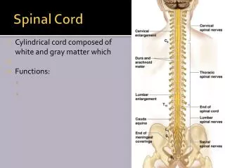
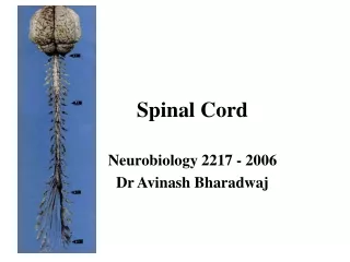
The Spinal Cord • Cylindrical in shape. • Occupies upper 2/3rd of the vertebral canal. • Begins superiorly at the Foramen magnum (Continuous with the Medulla Oblongata of Brain) • Ends inferiorly in adults at the Lower border of 1st Lumbar vertebra. • Surrounded by meninges & CSF.
The Spinal Cord • Fusiformly enlarged: • Cervical enlargement • Lumbar enlargement • Inferiorly tapers off into Conus Medullaris.
The Spinal Cord • Attached to coccyx by a Pia Mater extension (filum terminale).
The Spinal Cord Denticulate Ligament
Spinal Cord (Grooves or Sulcus) • Anterior median fissure • Posterior median sulcus
Spinal Cord (Internal Structure) • Gray matter in the center (H-shaped). • Posterior or Dorsal horns • Anterior or Ventral horns. • Lateral horn is only present in the thoracic, lumbar and sacral Regions. • The intermediate zone pierced by Central Canal.
Spinal Cord (Internal Structure) • White matter divided into: • Ventral Column or Funiculus • Dorsal Column or Funiculus • Lateral Column or Funiculus
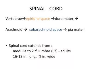
Meninges of Spinal Cord • Are continuous with the cranial meninges. • Dura mater • Arachnoid mater • Pia mater
Meninges of Spinal Cord • Epidural space: • Lies between vertebral column and dura mater • Contains blood vessels, areolar connective tissue & fat. • Subdural space: • Lies between the dura mater and arachnoid mater • Contains serous fluid. • Subarachnoid space: • Lies between arachnoid mater and Pia mater. • Contains cerebrospinal fluid (CSF) and blood vessels
The Spinal Cord • 8 Cervical nerves (C1-C8) • 12 Thoracic nerves (T1-T12) • 5 Lumbar nerves (L1–L5) • 5 Sacral nerves (S1–S5), • 1 Coccygeal nerve
The Spinal Cord • White matter • Myelinated axons • Divided into three columns (funiculi) • Ventral • Dorsal • lateral • Commissures: Connections between left and right halves • Gray with central canal in the center • White • Roots • Spinal nerves rootlets (dorsal and ventral roots). • Dorsal and ventral roots merge to form the spinal nerve.
Gray Matter • Consists of nerve cell bodies and their processes, neuroglia, and blood vessels • The nerve cells are multipolar and are of three main categories: • Sensory neurons: Receive impulses from the periphery of the body and whose axons constitute the ascending fasciculi of the white matter, are located in the dorsal horns • Motor neurons, which transmit impulses to the skeletal muscles, are located in the ventral horns (similar neurons in the lateral horn are the preganglionic neurons of the autonomic system) • Interneurons (Connector neurons) : linking sensory and motor neurons, at the same or different levels, which form spinal reflex arcs.