Download
1 / 37
410 likes | 527 Views
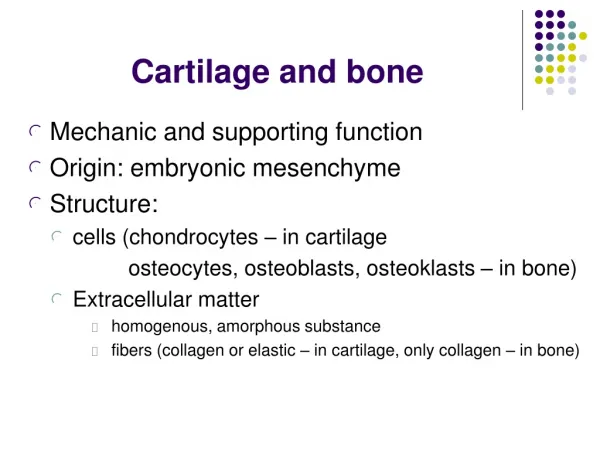
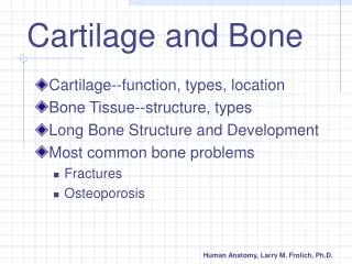
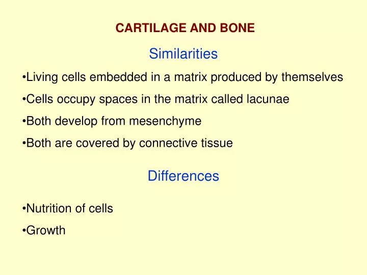
CARTILAGE AND BONE. Similarities. Living cells embedded in a matrix produced by themselves Cells occupy spaces in the matrix called lacunae Both develop from mesenchyme Both are covered by connective tissue. Differences. Nutrition of cells Growth.

E N D
CARTILAGE AND BONE Similarities • Living cells embedded in a matrix produced by themselves • Cells occupy spaces in the matrix called lacunae • Both develop from mesenchyme • Both are covered by connective tissue Differences • Nutrition of cells • Growth
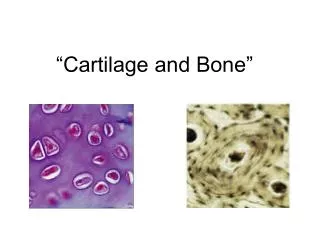
Histological preparation of hyalin cartilage territorial matrix perichondrium isogenous group Chondroblasts- appositional growth Chondrocytes - interstitial growth cell matrix lacunae Several growth factors : FGF, IGF, TGF-b and growth hormone influence cartilage growth
Condrocytes inside hyaline cartilage territorial matrix Isogenous group matrix
EM of chondrocyte and surrounding matrix proteoglycans territorial matrix collagen type II fibrils
piokl Proteoglycan aggregate
Cartilage matrix content • Water ≈ 70% • Organic components ≈ 30% (Collagen type II ≈ 40% and Proteoglycans ≈ 60%) • As a result the matrix has agel-like strucure that allows nutrition by diffusion and provides flexibility and mechanical resiliance • It also makes possible formation of isogenous groups by division of chondrocytes into the matrix and for interstitial growth of cartilage from inside
Types of cartilage • All of them contain collagen type II plus: • Hyalin cartilage ---- • Elastic cartilage elastic fibers • Articular cartilage collagen type I • Fibrocartilage collagen type I
Elasticcartilage perichondrium elastic fiber
Articular cartilage no perichondrium ! synovial cavity articular cartilage bone bone marrow
Fibrocartilage dense connective tissue collagen type I fibres rows of chondrocytes Found around nucleus pulposus of intervertebral disks. Again, no perichondrium !
Poor regeneration of cartilage • Except in young chidren, damaged cartilage regenerates with difficulties by activation of perichondrium. • If damage is extensive, perichondrium produces a scar of dense connective tissue
Bone BONE
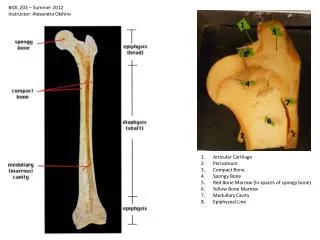
Longitudinal section of adult long bone Bone classification trabeculae/spicules cancelous/trabecular/spongy compact/cortical
Blood vessels in a system ofHaversian canals interconnected with Volkman’s canals 0steon - complex of concentric lamellae of bone surrounding a Haversian canal Cementingsubstance Interstitiallamellae endosteum periosteum Inner circumferential lamellae Outer circumferential lamellae
Thin section of calcified bone Osteon with Haversian canal Interstitial lamellae Lacunae of osteocytes Cement line
Histological preparation of decalcified bone Osteons with Haversian canals Volkman’s canal Inner circumferential lamellae Bone marrow
Primary, immature or woven bone (decalcified preparation) Spicule/trabecular (~0.2 mm) Beginning of formation of osteons, but well defined lamellae are not yet formed
CELLS OF BONE Mesenchymal cells differentiating into osteoblasts Capillary Not calcified bone matrix osteoid Osteocytes in lacunae with their processes interconnected Calcified bone matrix is penetrated by canaliculi
Calcification/mineralization of of bone matrix • Inorganic matter is ≈ 50% of the dry weight of bone matrix mainly in form of hydroxyapatite crystals - Ca10(PO4)6(OH)2 • Initial mineralization in woven bone or cartilage by matrix vesicles deposited by osteobalsts and chondrocytes and enriched in alkaline phosphatase and acidic phospholipids • Mineralization front - mineralization of matrix synthesized at already calcified mature bone surface • Deficiency of Ca and vitamin D negatively influence normal calcification process
osteocytes canaliculi calcified bone matrix osteoclast flat/resting osteoblasts blood vessel
ruffled border clear/adhesion zone
Howship’s lacunae osteoblasts making new bone
Remodeling Bone turnover and skeletal homeostasis In a young adult ≈ 30% of the total skeletal mass is renewed every year by remodeling in a balanced fashion Shfting the balance between bone formation and bone resorbtion - osteoporosis, osteopetrosis activation of osteoclasts Parathyroid hormone inhibition ofosteoclasts Calcitonin Calcification of new bone and surface recovery activation of osteoblasts osteoid
FORMATION OF HAVERSIAN CANAL Activation of osteoclasts Second concentric layer First concentric layer Inhibition of osteoclasts Activation of osteoblasts
Intramembranous bone formation New bone spicule/trabecular starts to grow Calcified matrix osteoid Process of remodeling starts Osteoblasts start to produce noncalcified bone matrix (osteoid) Activation of mesenchymal cells to turn into osteoblasts
Intramembranous formation of bones of the skull Active osteoblasts producing osteoid Calcified matrix New bone spicules/trabeculae growing
Intramembranous bone formation active osteoblasts producing osteoid osteoid osteocytes locked in bone matrix resting osteoblasts
1. Starting with cartilage model Endochondral bone formation 4. Invasion of osteoclasts followed by blood vessels and osteogenic cells 5. Beginning of new bone synthesis over the calcified cartilage matrix 6. Formation of primary center of ossification 7. Remodeling and growth 2. Formation of bony collar at the middle of diaphysis 3. Hypertrophy of chondrocytes followed by calcification of their marix and their death 8. Later a secondary center of ossification appears in the epiphyses that is separated from the primary center by growth plate of cartilage
Starting with cartilage model Hypertrophy and degeneration of chondrocytes
Bony collar on the surface at the middle of diaphysisformed by intramembranous bone formation Beginning of formation of primary center of ossification
Propagation of the process of primary ossificationfollowed by remodeling of the newly formed bone trabeculae Closer look at this area next At the same time formation of bony collar also continues by Intramembranous bone formation
Columns of dividing chondrocytes - interstitial growth Hypertrophy of chondrocytes Calcification of cartilage matrix and death of chondrocytes Invasion of blood vessels and osteogenic cells and beginning of bone formation over calcified cartilage matrix Remodelingstarts
Secondary center of ossification Epiphysis Metaphysis Growth/epiphysial plate Diaphysis Advancing of primary ossification
Endochondral bone formation 1. Starting with cartilage model 2. Formation of bony collar on the surface at the middle of diaphysis 3. Hypertrophy and degeneration of chondrocytes and calcification of their marix 4. Invasion of osteoclasts followed by blood vessels and osteogenic cells 5. Beginning of new bone synthesis over the calcified cartilage matrix 6. Formation of primary center of ossification 7. Remodeling and growth 8. Later a secondary center of ossification appears in the epiphysis