Download
1 / 19
200 likes | 234 Views
CARTILAGE & BONE. Objectives: By the end of this lecture, the student should describe the microscopic structure, distribution and growth of the different types of: (1) Cartilage. (2) Bone. CARTILAGE. Cartilage is a specialized type of C.T. with a rigid matrix.

E N D
CARTILAGE & BONE Objectives: • By the end of this lecture, the student should describe the microscopic structure, distribution and growth of the different types of: (1) Cartilage. (2) Bone.
CARTILAGE • Cartilage is a specialized type of C.T. with a rigid matrix. • Cartilage is usuallynonvascular (avascular). • 3 Types: • Hyaline cartilage. • Elastic cartilage. • Fibrocartilage.
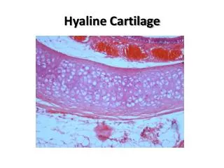
Hyaline Cartilage 1- Perichondrium: • Vascular C.T. membrane formed of 2 layers: • Outer fibrous layer:dense fibrous C.T. • Inner chondrogenic layer:contains chondroblasts ( no lacunae). They secrete cartilage matrix and give rise to chondrocytes.
Hyaline Cartilage 2- Cells (Chondrocytes): • Found in spaces called lacunae. • Young chondrocytes: are small & present singly in their lacunae. • Mature chondrocytes: are large, and are found singly or in groups of 2, 4 or 6 cells in their lacunae (cell nests). 3- Matrix: • Homogeneous and basophilic. • Contains collagen type II.
Hyaline Cartilage • Sites of hyaline cartilage: • Foetal skeleton. • Costal cartilages. • Articular surfaces of bones. • Nose, trachea & bronchi.
Elastic Cartilage • Similar to hyaline cartilage + elastic fibres in the matrix. • Sites: • External ear. • Epiglottis.
Fibrocartilage • No perichondrium. • Rows of chondrocytes in lacunae separated by parallel bundles of collagen fibers (type I). • Sites: e.g. Intervertebral disks.
Growth of cartilage 1. Appositional growth: • Is produced by the activity of Chondroblasts in the inner chondrogenic layer. • It leads to increase in width. 2. Interstitial growth: • Is produced by division and activity of mature chondrocytes. • It leads to increase inlength.
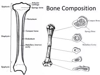
BONE • Bone is a specialized type of C.T. with a hard matrix. • Types: 2 types • Compact and spongy (cancellous( bone. • Components: • Bone Cells: 4 types. • Bone Matrix (calcified osteoid tissue): hard because it is calcified (Calcium salts). It contains type I collagenfibers. It forms bone lamellae and trabeculae. • Periosteum. • Endosteum. • Functions: • body support. • protection of vital organs as brain & bone marrow. • calcium store.
Bone Cells 1- Osteogenic Cells: • in periosteum & endosteum. • Fate: give rise to osteoblasts. 2- Osteoblasts: • in periosteum & endosteum. • Origin: osteogenic cells. • Function: They secrete the bone matrix & deposit Ca salts in it. • Fate: change to osteocytes.
Bone Cells 3- Osteocytes: • Branched cells. • Present singly in lacunae. Their branches run in the canaliculi. • Origin: osteoblasts. • Function: They maintain the bone matrix.
Bone Cells 4- Osteoclasts: • Large multinucleated cells on bony surfaces, in Howship’s lacunae. • They have striated or ruffled border. • Cytoplasm is rich in lysosomes. • Origin: blood monocytes. • Function: bone resorption.
Compact Bone • It is found in the diaphysis of long bones. • Consists of: 1- Periosteum: • Outer fibrous layer. • Inner osteogenic layer. 2- Endosteum. 3- Bone Lamellae. 4- Bone Cells.
Compact Bone Bone Lamellae: 1- Haversian Systems (Osteons): • Longitudinal cylinders. • Each is formed of concentric bone lamellae & a Haversian canal, running in the center. • Volkmann’s canals:connect the Haversian canals together. They run obliquely or transversely. 2. External Circumferential Lamellae. 3- Internal Circumferential Lamellae. 4- Interstitial Lamellae: between osteons.
Spongy (Cancellous) Bone • In flat bones & epiphysis of of long bones. • Consists of : • Periosteum. • Endosteum. • Irregular bone trabeculae. • Many irregular bone marrow spaces. • Bone Cells. • No Haversian systems (no osteons).
Growth of Bone • Appositional growth: • Is produced by the activity of osteoblasts. • It leads to increase in width. • Growth in length: • Is produced by the activity of epiphyseal plate of cartilage.