Download
1 / 50
500 likes | 639 Views
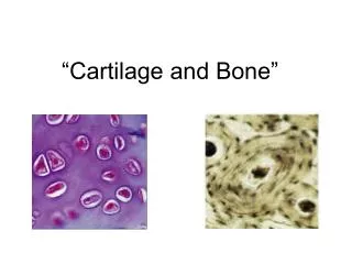
Cartilage and bone. Mechanic and supporting function Origin: embryonic mesenchyme Structure: cells (chondrocytes – in cartilage osteocytes, osteoblasts, osteoklasts – in bone) Extracellular matter homogenous, amorphous substance

E N D
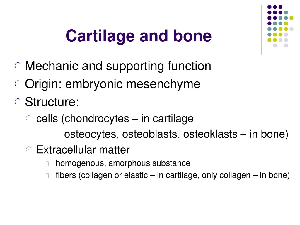
Cartilage and bone • Mechanic and supporting function • Origin: embryonic mesenchyme • Structure: • cells (chondrocytes – in cartilage osteocytes, osteoblasts, osteoklasts – in bone) • Extracellular matter • homogenous, amorphous substance • fibers (collagen or elastic – in cartilage, only collagen – in bone)
Cartilage properties • Avascular tissue, without nerves, • Decreased reparative ability, regeneration from perichondrium • Perichondrium – dense collagen c. t. attached to cartilage • inner chondrogenic layer • outer c. t. layer
Cell of cartilage • chondroblasts • immature chondrocytes in inner layer of perichondrium • chondrocytes – in cartilage • basofilic cells → proteosynthesis, mitochondria, GER, Golgi apparatus, cytoskeleton. • produce amorphous matrix and fibers.
Chondrocytes – isogenous groups,– lacunae, – basophilic capsule of teritorial matrix • Isogenous group + adjacent area of extra- cellular matter (territorial matter) = chondron
Collagen II or I fibers Elastic fibers Glykosaminoglycans –hyaluronic acid, chondroitin-sulphate Proteoglycans Glykoproteins Extracellular matter
Types of cartilage • Hyaline(hyalos=glass) – the most frequent, precursor of many bones in skeleton, covers articular surfaces, forms part of ribs skeleton of the nose, trachea, larynx • Elastic - auricula, tuba auditiva, larynx, epiglottis • Fibrocartilage - intervertebral discs, symphysis pubis, articular discs and meniscus
Elastic cartilage • elastic fibers in amorphous matrix; special staining: resorcin, fuchsin and orcein. • Chondrocytes
Fibrocartilage • Chondrocytes • Thick bundles of collagenous fibers • matrix • without perichondrium
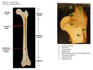
Bone • specialized form of c. t. Bone tissue structure • Bone cells • Extracellular matrix • Organic – colagenos fibers – amorphous matrix • Inorganic – minerals • Periosteum
Bone tissue • Periosteum – covers outer surface of bone: • Inner layer (osteoblasts, fibroblasts) • outer layer (only fibroblasts) Periosteum is attached by Sharpey‘s fibers. • Endosteum – membrane with one layer of cells (osteoblasts, osteoclasts), covers inner surface of bone turned to the bone cavity)
Bone cells • osteoprogenitor cells – stem cells, in periosteum a endosteum • osteoblasts produce organic matter, and transform into osteocytes
Osteocyte • Inlakuna, • Numerous processes in canaliculi ossium (cell communication)
Osteoclasts • large cells (up to 100 µm), polynucleated cells (up to 50 N), arrise by fussion of monocytes • in Howship‘s lakuna • lysosomal enzymes digest collagennous fibers
Bone matrix • collagen fibers – collagen I (cca 90 % of org. matter) • amorphous matrix – osteoid. • strength of matrix is caused by content of inorganic salts (hydroxyapatite).
Types of bone tissue • 2 types: /according to arrangement of collagen fibers/ • Fibrillar (woven) bone– primary • Lamellar bone– secondary • compact – wall of long bone diaphysis, surface layer of epiphysis • spongy /trabecular/ – inner part of epiphysis
Lamellar bone • Lamellae – thin plates with regularly arranged collagen fibers • Haversian systems - osteons • Circumferential lamellae • outer • inner • Interstitial lamellae
Lamellar bone Haversian and Volkmann‘s canals
Lamellar bone – spongy type • Matrix is also organized into the lamellae, but dont formHaversian systems.
Histogenesis of bone tissue • Endochondral ossification – hyaline cartilage is model for bone (long bones) • Intramembranous ossification – mesenchyme membrane is model, mesenchymocetes differentiate into osteoblasts (skull bone, part of mandibule and clavicula)
Periosteal bone collar Undifferentiated cells in the perichondrium become osteoblasts, and the perichondrium is now the periosteum.
Chondrocytes at the center of the growing (proliferating zone) cartilage model enlarge (zone ofhypertrophy). The matrix calcifies(zone of calcification) and chondrocytes die. The rest of matrix form trabecular processes – spicules. Blood vessels penetrate cartilage and carry the osteoblasts from periosteum. Osteoblasts cover the spicules and produce osteoid (ossification zone). Ossification spreads in long axis of bone.
Osteoclasts resorb primitive bone (zone of resorption) and medullary cavity is formed. Simillar process begins in epiphyses. Ossification spread radially. Also epiphyseal plates ossify at the end of body growth (cca 18 years).
Endochondral ossification • Zone of normal cartilage • Zone of proliferated cartilage • Zone of hypertrophic cartilage • Zone of calcification • Line of erosion • Zone of ossification • Zone of resorption
Bone growth • cannot occur interstitally as cartilage growth does, because its rigid, mineralized matrix traps osteocytes and prevents them from dividing mitotically. Growth occurs in two directions: • in length, by maintenance and growth of epiphyseal plate of cartilage. These plates allow a bone to expand lengthwise. • in diameter, by continuous formation of bone around the periphery of the diaphysis.
Endochondral ossification Normal hyaline cartilage Proliferating cartilage (growth) Hypertrophic cartilage Calcified cartilage - calcified matrix LINE of EROSION spicules blood vessels osteoid osteoblasts B O N E