Download
1 / 69
690 likes | 742 Views
BONE & CARTILAGE-1. This resource is licensed under the Creative Commons Attribution Non-Commercial & No Derivative Works License. Objectives For both Bone & Cartilage sessions. Describe the microscopic appearance of the different histological types of cartilage.

E N D
BONE & CARTILAGE-1 This resource is licensed under the Creative Commons Attribution Non-Commercial & No Derivative Works License
Objectives For both Bone & Cartilage sessions Describe the microscopic appearance of the different histological types of cartilage. Identify the cell types found in bone and describe the microscopic organisation of cortical and trabecular bone. Consolidate the information on bone development gained in lectures by observing the microscopic appearance of developing bones.
SLIDE 64 Trachea (chick) Hyaline cartilage At low magnification determine whether the section is transverse or longitudinal? Identify the position of the hyaline cartilage. 1.0 mm
SLIDE 64 Trachea (chick) Hyaline cartilage fold in section At low magnification determine whether the section is transverse or longitudinal? Answer : transverse section of trachea. Identify the position of the hyaline cartilage. muscle lumen of trachea C shaped hyaline cartilage lining epithelium of trachea 1.0 mm
SLIDE 64 Trachea (chick) Hyaline cartilage What is the function of the hyaline cartilage of the trachea? 250 µm
SLIDE 64 Trachea (chick) Hyaline cartilage lumen perichondrium What is the function of the hyaline cartilage of the trachea? It maintains the shape of the trachea. It resists compression; but allows movement due to the C shaped rings. It provides a resilient support to the trachea. cartilage tracheal epithelium 250 µm
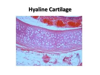
SLIDE 64 Trachea (chick) Hyaline cartilage Identify : perichondrium. young and mature chondrocytes. chondrocyte lacunae and matrix. 100 µm
SLIDE 64 Trachea (chick) Hyaline cartilage perichondrium matrix Identify : perichondrium. young and mature chondrocytes. chondrocyte lacunae and matrix. chondrocyte lacuna young chondrocyte mature chondrocytes 100 µm
SLIDE 64 Trachea (chick) Hyaline cartilage Higher magnification area from previous slide. Perichondrium covers both surfaces and is composed of an outer fibrous layer of connective tissue and an inner layer of chondrogenic cells. 25 µm
SLIDE 64 Trachea (chick) Hyaline cartilage matrix chondrocytes in lacunae Higher magnification area from previous slide. Perichondrium covers both surfaces and is composed of an outer fibrous layer of connective tissue and an inner layer of chondrogenic cells. perichondrium 25 µm
SLIDE 126 Articular cartilage Hyaline cartilage Whole section. Again determine whether the section is transverse or longitudinal? 1 cm
SLIDE 126 Articular cartilage Hyaline cartilage metaphysis diaphysis epiphysis articular cartilage epiphysial growth plate Whole section. Again determine whether the section is transverse or longitudinal? Answer : Longitudinal section of bone. 1 cm
SLIDE 126 Articular cartilage Hyaline cartilage What is the function of the hyaline cartilage of the articular surface? 25 µm
What is the function of the hyaline cartilage of the articular surface? Friction reduction; provides a smooth sliding surface for joint movement. Again provides a resilient support and resists compression. SLIDE 126 Articular cartilage Hyaline cartilage hyaline cartilage articular surface 25 µm
SLIDE 126 Articular cartilage Hyaline cartilage Enlarged area from previous slide. Chondrocytes now visible. Distal Femur (Dog) Hyaline cartilage of femoral condyles and trochlea. articular surface 100 µm
SLIDE 126 Articular cartilage Hyaline cartilage Is a perichondrium present in articular hyaline cartilage? 50 µm
SLIDE 126 Articular cartilage Hyaline cartilage flattened chondrocytes Is a perichondrium present in articular hyaline cartilage? No. Note the flattened appearance of chondrocytes at and close to the articulating surface. multiple chondrocytes in single lacunae matrix articular surface 50 µm
SLIDE 126 Articular cartilage Hyaline cartilage Perichondrium covers all hyaline cartilage except that forming articular surfaces in joints. cartilage matrix mature chondrocytes in lacunae 25 µm
Articular cartilage Hyaline cartilage 1. What cells are responsible for the synthesis of the cartilage matrix? . 2. Name the predominant components of the extracellular matrix of hyaline cartilage? . What sub-cellular organelles would you expect to find in cells actively involved in the synthesis of this matrix? . Does cartilage contain blood vessels? .
Articular cartilage Hyaline cartilage 1. What cells are responsible for the synthesis of the cartilage matrix? Chondrocytes. 2. Name the predominant components of the extracellular matrix of hyaline cartilage? . What sub-cellular organelles would you expect to find in cells actively involved in the synthesis of this matrix? . Does cartilage contain blood vessels? .
Articular cartilage Hyaline cartilage 1. What cells are responsible for the synthesis of the cartilage matrix? Chondrocytes. 2. Name the predominant components of the extracellular matrix of hyaline cartilage? Thin fibrils of collagen type II. Amorphous ground substance : Proteoglycans. Chondronectin. Water. Hyaluronic acid. What sub-cellular organelles would you expect to find in cells actively involved in the synthesis of this matrix? . Does cartilage contain blood vessels? .
Articular cartilage Hyaline cartilage 1. What cells are responsible for the synthesis of the cartilage matrix? Chondrocytes. 2. Name the predominant components of the extracellular matrix of hyaline cartilage? Thin fibrils of collagen type II. Amorphous ground substance : Proteoglycans. Chondronectin. Water. Hyaluronic acid. What sub-cellular organelles would you expect to find in cells actively involved in the synthesis of this matrix? Abundant rough endoplasmic reticulum (RER) and well developed golgi. Does cartilage contain blood vessels? .
Articular cartilage Hyaline cartilage 1. What cells are responsible for the synthesis of the cartilage matrix? Chondrocytes. 2. Name the predominant components of the extracellular matrix of hyaline cartilage? Thin fibrils of collagen type II. Amorphous ground substance : Proteoglycans. Chondronectin. Water. Hyaluronic acid. What sub-cellular organelles would you expect to find in cells actively involved in the synthesis of this matrix? Abundant rough endoplasmic reticulum (RER) and well developed golgi. Does cartilage contain blood vessels? No.
SLIDE 128 Epiglottis (cat) Elastic cartilage Low magnification view of the epiglottis. 0.5 mm Dog (eviscerated preparation, dorsal view) T – tongue E – epiglottis A – arytenoid cartilage V – vestibulum esophagi* L – limen pharyngoesophageum* (* pars laryngea pharyngis) O – oesophagus (opened) Tr – trachea (opened)
SLIDE 128 Epiglottis (cat) Elastic cartilage folds in section Low magnification view of the epiglottis. Elastic fibres are stained black in this preparation. 0.5 mm
SLIDE 128 Epiglottis (cat) Elastic cartilage Do the elastic fibres have a regular or irregular arrangement? 100 µm
Do the elastic fibres have a regular or irregular arrangement? Mainly irregular : a dense network of branching fibres around the chondrocytes, less dense towards the perichondrium. SLIDE 128 Epiglottis (cat) Elastic cartilage chondrocytes Do the elastic fibres have a regular or irregular arrangement? elastic fibres stained black 100 µm
Do the elastic fibres have a regular or irregular arrangement? Mainly irregular : a dense network of branching fibres around the chondrocytes, less dense towards the perichondrium. How can you relate this to the function of the epiglottis? SLIDE 128 Epiglottis (cat) Elastic cartilage chondrocytes Do the elastic fibres have a regular or irregular arrangement? elastic fibres stained black 100 µm
Do the elastic fibres have a regular or irregular arrangement? Mainly irregular : a dense network of branching fibres around the chondrocytes, less dense towards the perichondrium. How can you relate this to the function of the epiglottis? The epiglottis can return to its normal shape after distortion. SLIDE 128 Epiglottis (cat) Elastic cartilage chondrocytes Do the elastic fibres have a regular or irregular arrangement? elastic fibres stained black 100 µm
SLIDE 128 Epiglottis (cat) Elastic cartilage chondrocyte in lacuna Elastic cartilage is found in areas which need to resist mechanical deformation. As well as the epiglottis it can be found in the larynx, external auditory canals and auditory tubes. elastic fibres stained black matrix 25 µm
SLIDE 130 Intervertebral disc(mouse) Fibrocartilage Whole section 1.0 mm
SLIDE 130 Intervertebral disc(mouse) Fibrocartilage Selected area x1 objective 1 mm
SLIDE 130 Intervertebral disc(mouse) Fibrocartilage intervertebral discs Selected area x1 objective V : bones of vertebrae V V V muscle 1 mm
SLIDE 130 Intervertebral disc(mouse) Fibrocartilage Selected area x4 objective 250 µm
SLIDE 130 Intervertebral disc(mouse) Fibrocartilage endplates bone Selected area x4 objective nucleus pulposus annulus fibrosus 250 µm
SLIDE 130 Intervertebral disc(mouse) Fibrocartilage Observe the predominant concentric collagen fibre layers in the annulus. Explain what you understand the term ‘slipped disc’ to mean. 100 µm
SLIDE 130 Intervertebral disc(mouse) Fibrocartilage nucleus pulposus bone of vertebra Observe the predominant concentric collagen fibre layers in the annulus. Explain what you understand the term ‘slipped disc’ to mean. Displacement of the nucleus pulposus annulus fibrosus 100 µm
SLIDE 130 Intervertebral disc(mouse) Fibrocartilage concentric rings of annulus fibrosus What collagen type predominates in the Annulus Fibrosus? 50 µm
SLIDE 130 Intervertebral disc(mouse) Fibrocartilage concentric rings of annulus fibrosus What collagen type predominates in the Annulus Fibrosus? Collagen type I fibres. 50 µm
Changes to the inter-vertebral disc induced by axial compression and bending. Which sites in 3 are experiencing extension or tension? bone endplate annulus fibrosus nucleus pulposus 2 1 3
Changes to the inter-vertebral disc induced by axial compression and bending. Which sites in 3 are experiencing extension or tension? bone endplate annulus fibrosus compression extension nucleus pulposus 2 1 3
Consider the compositional similarities and differences between the three distinct types of cartilage examined.
SLIDE 133 Skull(cat)Cancellous (spongy or trabecular) bone Whole section. Consider how a section through a bath sponge might appear! 5.0 mm
SLIDE 133 Skull(cat)Cancellous (spongy or trabecular) bone Selected area. Identify : Bone marrow between trabeculae. Bone matrix. Osteocytes within the bone matrix. Osteoblasts on forming surfaces. Lining cells : line vascular canals and marrow spaces. 250 µm
SLIDE 133 Skull(cat)Cancellous (spongy or trabecular) bone lining cells bone marrow Selected area. Identify : Bone marrow between trabeculae. Bone matrix. Osteocytes within the bone matrix. Osteoblasts on forming surfaces. Lining cells : line vascular canals and marrow spaces. trabeculae bone matrix osteoblasts osteocytes in matrix 250 µm
SLIDE 133 Skull(cat)Cancellous (spongy or trabecular) bone Identify osteoclasts if present. 50 µm
SLIDE 133 Skull(cat)Cancellous (spongy or trabecular) bone Identify osteoclasts if present. Which of these cells is responsible for forming bone? osteoclast 50 µm
SLIDE 133 Skull(cat)Cancellous (spongy or trabecular) bone O : osteoblasts bone matrix O Identify osteoclasts if present. Which of these cells is responsible for forming bone? Osteoblasts synthesize the organic matrix. The secretory surface faces the bone. Which of these cells is responsible for removing bone? osteoclast 50 µm
SLIDE 133 Skull(cat)Cancellous (spongy or trabecular) bone O : osteoblasts bone matrix O Identify osteoclasts if present. Which of these cells is responsible for forming bone? Osteoblasts synthesize the organic matrix. The secretory surface faces the bone. Which of these cells is responsible for removing bone? Osteoclasts. bone marrow osteoclast osteocytes 50 µm
SLIDE 133 Skull(cat)Cancellous (spongy or trabecular) bone Active osteoclasts contain many lysosomes and facing the bone have a folded surface termed a ‘ruffled border’. From which cells are osteoclasts derived and describe one characteristic feature of mature osteoclasts? osteoclast 50 µm