Download
1 / 25
250 likes | 288 Views
CARTILAGE & BONE. Objectives: By the end of this lecture, the student should describe the microscopic structure, distribution and growth of the different types of: (1) Cartilage. (2) Bone. CARTILAGE. Cartilage is a specialized type of C.T. with a rigid matrix.

E N D
CARTILAGE & BONE • Objectives: • By the end of this lecture, the student should describe the microscopic structure, distribution and growth of the different types of: (1) Cartilage. (2) Bone.
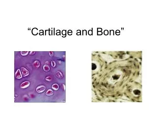
CARTILAGE • Cartilage is a specialized type of C.T. with a rigid matrix. • Cartilage is usually nonvascular (avascular). • 3 Types: • Hyaline cartilage. • Elastic cartilage. • Fibrocartilage.
Hyaline Cartilage 1- Perichondrium: • Vascular C.T. membrane formed of 2 layers: • Outer fibrous layer:dense fibrous C.T. • Inner chondrogenic layer:contains chondroblasts ( no lacunae). They secrete cartilage matrix and give rise to chondrocytes.
Hyaline Cartilage 2- Cells (Chondrocytes): • Found in spaces called lacunae. • Young chondrocytes: are small & present singly in their lacunae. • Mature chondrocytes: are large, and are found singly or in groups of 2, 4 or 6 cells in their lacunae (cell nests). 3- Matrix: • Homogeneous and basophilic. • Contains collagen type II.
Hyaline Cartilage • Sites of hyaline cartilage: • Foetal skeleton. • Costal cartilages. • Articular surfaces of bones. • Nose, trachea & bronchi.
Growth of cartilage 1. Appositional growth: • Is produced by the activity of Chondroblasts in the inner chondrogenic layer. • It leads to increase in width. 2. Interstitial growth: • Is produced by division and activity of mature chondrocytes. • It leads to increase inlength.
Elastic Cartilage • Similar to hyaline cartilage + elastic fibres in the matrix. • Sites: • External ear. • Epiglottis.
Fibrocartilage • No perichondrium. • Rows of chondrocytes in lacunae separated by parallel bundles of collagen fibers (type I). • Sites: e.g. Intervertebral disks.
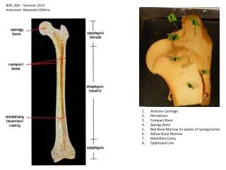
BONE • Bone is a specialized type of C.T. with a hard matrix. • Types: 2 types • Compact and spongy (cancellous( bone. • Components: • Bone Cells: 4 types. • Bone Matrix: hard because it is calcified (Calcium salts). It contains type I collagen fibers. It forms bone lamellae and trabeculae. • Periosteum. • Endosteum. • Functions: • body support. • protection of vital organs as brain & bone marrow. • calcium store.
Bone Cells 1- Osteogenic Cells: • in periosteum & endosteum. • Fate: give rise to osteoblasts. 2- Osteoblasts: • in periosteum & endosteum. • Origin: osteogenic cells. • Function: They secrete the bone matrix & deposit Ca salts in it. • Fate: change to osteocytes.
Bone Cells 3- Osteocytes: • Branched cells. • Present singly in lacunae. Their branches run in the canaliculi. • Origin: osteoblasts. • Function: They maintain the bone matrix.
Bone Cells 4- Osteoclasts: • Large multinucleated cells on bony surfaces, in Howship’s lacunae. • They have striated or ruffled border. • Cytoplasm is rich in lysosomes. • Origin: blood monocytes. • Function: bone resorption.
Compact Bone • It is found in the diaphysis of long bones. • Consists of: 1- Periosteum: • Outer fibrous layer. • Inner osteogenic layer. 2- Endosteum. 3- Bone Lamellae. 4- Bone Cells.
Compact Bone Bone Lamellae: 1- Haversian Systems (Osteons): • Longitudinal cylinders. • Each is formed of concentric bone lamellae & a Haversian canal, running in the center. • Volkmann’s canals:connect the Haversian canals together. They run obliquely or transversely. 2. External Circumferential Lamellae. 3- Internal Circumferential Lamellae. 4- Interstitial Lamellae: between osteons.
Spongy (Cancellous) Bone • In flat bones & epiphysis of of long bones. • Consists of : • Periosteum. • Endosteum. • Irregular bone trabeculae. • Many irregular bone marrow spaces. • Bone Cells. • No Haversian systems (no osteons).
Growth of Bone • Appositional growth: • Is produced by the activity of osteoblasts. • It leads to increase in width. • Growth in length: • Is produced by the activity of epiphyseal plate of cartilage.
Clinical application Disk Prolapse • It occurs more often on the posterior portion of the intervertebral disks, particularly in the lumbar region. • Tear or break in the laminae of the annulus fibrousus, through which the gel-like nucleus pulposus extrudes. • The disk may dislocate or slip compresses the lower spinal nerves leading to severe pain in the lower back and the lower limbs.
Clinical application Serum (blood) alkaline phosphatase It is an indicator of bone formation----why? Osteobalsts are rich in alkaline phosphatase enzyme.
Clinical application Osteopetrosis It is a genetic disorder where osteoclasts do not possess a ruffled border these cells can not resorb bone Effects: • Increased bone density. • Anemia (may be----why?)
Clinical application Osteoporosis It is related to decreasing bone mass, which becomes more serious after menopause, where estrogen secretion drops appreciably. Pathogenesis: Binding of estrogen to specific receptors on Osteobalstsactivate the cells to synthesize and secrete bone matrix. With diminished secretion of estrogen, osteoclasts’ activity (bone-resorption) will be greater than bone deposition reducing bone mass to the extent at which the bone cannot withstand stress and break easily.
Clinical application Rickets It is a disease in infants and children who are deficient in vitamin D. Pathogenesis: Deficiency of vitamin D intestinal mucosa cannot absorb calcium from diet disturbances in ossification of the epiphyseal cartilages and disorientation of the cells in the metaphysis poorly calcified bone matrix
Clinical application Osteomalacia (adult Rickets) It results from deficiency of vitamin D. The newly formed bone in the process of remodeling does not calcify properly. This condition becomes severe during pregnancy because the fetus requires calcium, which must be supplied by the mother.