Download
1 / 32
320 likes | 484 Views
Spinal Cord and Reflexes. http://biology.clc.uc.edu. Much of the text material is from, “Principles of Anatomy and Physiology, 14th edition” by Gerald J. Tortora and Bryan Derrickson (2014). I don’t claim authorship. Other sources are noted when they are used.

E N D
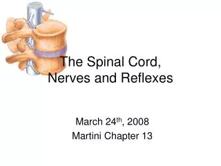
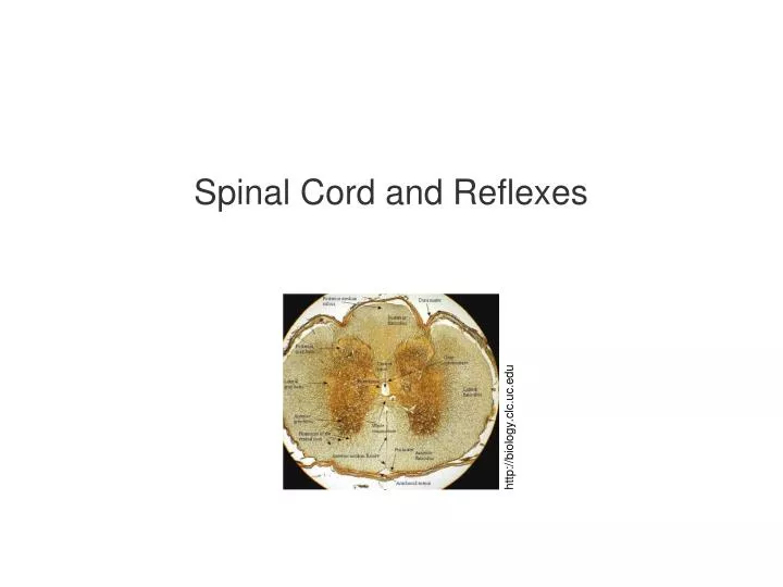
Spinal Cord and Reflexes http://biology.clc.uc.edu
Much of the text material is from, “Principles of Anatomy and Physiology, 14th edition” by Gerald J. Tortora and Bryan Derrickson (2014). I don’t claim authorship. Other sources are noted when they are used. Mappings of the lecture slides to the 12th and 13th editions are provided in the supplements.
Outline • Functional anatomy • Spinal reflexes • Poliomyelitis
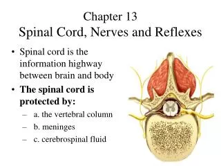
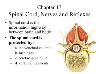
Vertebral Column • The spinal cord is located within the vertebral canal of the vertebral column. • The foramina of the vertebrae stacked on top of each other form the vertebral canal. • The vertebrae provides structure and a protective enclosure for the spinal cord. • The vertebral ligaments, meninges, and cerebrospinal fluid provide additional protection. Foramina (plural) = openings in bones. Figure 13.1 Chapter 13, page 443
Vertebral Column (continued) http://media.wiley.com
Spinal Nerves • The spinal nerves, and the nerves that branch from them, are part of the peripheral nervous system (PNS). • They connect the CNS to sensory receptors, muscles, and glands. • The 31 pairs of spinal nerves are named and numbered according to the segment of the vertebral column from which they emerge. Figure 13.2 Chapter 13, page 446
Spinal Nerves (continued) Spinal nerve http://www.csus.edu
Segments • Eight pairs of cervical nerves, C1 through C8 • Twelve pairs of thoracic nerves, T1 through T12 • Five pairs of lumbar nerves, L1 through L5 • Five pairs of sacral nerves, S1 through S5 • One pair of cocygeal nerves, Co1 Figure 13.2 Chapter 13, page 446
Anterior and Posterior Roots • Two bundles of axons, called roots, connect each spinal nerve to a segment of the spinal cord by even smaller bundles of axons known as rootlets. • The posterior root and its rootlets contain sensory axons to conduct nerve impulses from the sensory receptors in the skin, muscles, and internal organs to the CNS. • Each posterior root has a swelling, called a posterior root ganglion, that contains the cell bodies of sensory neurons. • The anterior root and its rootlets consist of motor axons to conduct nerves impulses from the CNS to skeletal muscles. Nerve impulse = action potential. Figure 13.3 Chapter 13, page 446
Anterior and Posterior Roots (continued) Posterior root and its ganglion (sensory) Anterior root (motor) http://www.csus.edu
Spinal Cord Section • A freshly-dissected section of spinal cord displays white matter and an inner core of gray matter. • A transverse section of the spinal cord and its features are shown in the illustration. Figure 13.3 Chapter 13, page 447
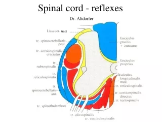
Spinal Cord Section (continued) • Meninges • Central canal • Anterior median fissure • White matter • Gray matter • Posterior horn • Anterior horn • Lateral horn • Fiber tracts • Commissural fibers http://biology.clc.uc.edu
Gray Matter • The posterior gray horns contain axons of incoming sensory neurons, and cell bodies and axons of interneurons. • The anterior gray horns contain somatic motor nuclei—clusters of cell bodies of somatic motor neurons that control the contraction of skeletal muscles. • The lateral gray horns, found only in the thoracic and lumbar segments, contain motor nuclei for the autonomic nervous system. • These nuclei control the activity of smooth muscle, and endocrine and exocrine glands. Figure 13.3 Chapter 13, page 447
White Matter • White matter is organized in columns in the spinal cord. • Each column consists of bundles of myelinated axons that have either a common origin or destination and carry similar types of information. • The bundles, which can extend long distances in the spinal cord, are known as fiber tracts. Figure 13.3 Chapter 13, page 447
White Matter (continued) • Ascending or afferent tracts consist of axons that convey sensory information to the brain. • Descending or efferent tracts have axons that convey motor infor-mation from the brain. • The ascending and descending tracts are continuous and uninter-rupted by synapses. • This arrangement helps increase the transmission speed of nerve impulses that must travel long distances since there are no inter-mediate synaptic delays. Figure 13.3 Chapter 13, page 447
Fiber Tracts http://www.hopkins-arthritis.org
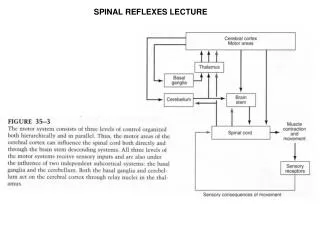
Review—Input and Output • Sensory input is conveyed from sensory receptors to the posterior horns of the spinal cord. • Motor output is conveyed from the anterior and lateral horns to effectors (muscles and glands). • The sensory and motor pathways are shown in the textbook illustration. Figure 13.4 Chapter 13, page 448
Dermatomes • Somatic sensory neurons or touch, vibration, temperature, and pain carry nerve impulses from the skin to the spinal cord and then to the brain. • Each spinal nerve consist of sensory neurons that serve a segment of the body. • The area of the skin that provides input to the CNS via one pair of spinal nerves or the trigeminal (V) nerve is known as a dermatome. • The nerve supply in adjacent dermatomes overlaps to some degree, so there is some redundancy. Figure 13.11 Chapter 13, page 460
Dermatomes (continued) Anterior view Posterior view medical-dictionary.thefreedictionary.com
Injury Location • Knowledge of which spinal cord segments supply each dermatome makes it possible to locate damaged regions of the spinal cord. • If the skin of a specific region is stimulated, but sensations are not perceived, the spinal cord segments supplying that dermatome may be damaged. • In areas where the overlap in dermatomes is substantial, very little loss of sensation may occur if one of the nerves supplying the der-matome is not damaged. • Information about innervation patterns of spinal nerves is also used in surgical procedures and anesthesia. Chapter 13, page 460
Reflexes • A reflex is a rapid, automatic response involving a sequence of physio-logical actions in response to a stimulus. • Some reflexes are inborn, such as pulling one’s hand away from a hot stove. • Higher-level reflexes may be learned, such as those involved in driving an automobile. Chapter 13, page 462
Reflexes (continued) • A spinal reflex involves sensory-motor integration in the spinal cord instead of the brain. • An example is the patellar (knee jerk) reflex mediated by the spinal cord. • A cranial reflex is when the integration occurs in the brainstem or at a higher level. • An example is the saccadic tracking movements of the eyes when reading a book. Chapter 13, page 462
Types of Reflexes • Somatic reflexes involve the contraction of one or more skeletal mus-cles. • Autonomic reflexes involve smooth muscle, cardiac muscle, or glands. • These reflexes are controlled by the sympathetic and parasympathetic divisions of the autonomic nervous system, which we will discuss in an upcoming lecture. Chapter 13, page 462
Reflex Arcs • The pathway of nerve impulses in a reflex is known as a reflex arc. • The functional components of a reflex arc consist of: • Sensory receptor—production of generator (graded) electrical potentials. • Sensory neuron—propagation of nerve impulses to the spinal cord. • Integrating center—monosynaptic (no interneuron) or polysyn-aptic (one or more interneurons). • Motor neuron—propagation of nerve impulses from the spinal cord to an effector. • Effector—response of the muscle or gland. Figure 13.13 Chapter 13, page 462
A Few Somatic Reflexes • Stretch reflex—causes contraction of skeletal muscles in response to stretching of a skeletal muscle. • Tendon reflex—controls skeletal muscle tension by causing muscle relaxation before the muscle force becomes too great that tendons could be damaged. • Flexor reflex—causes contraction of skeletal muscle in response to, for example, a painful stimulus. • Cross-extensor reflex—along with some flexor reflexes, causes con-traction of skeletal muscles on the opposite side of the body to main-tain balance. Figures 13.14 through 3.16 Chapter 13, page 463
Poliomyelitis • Poliomyelitis (or just, polio) is caused by the poliovirus, an RNA virus of about 7500 nucleotides. • Its initial symptoms include fever, severe headache, stiff neck and back, muscle pain and weakness, and loss of some somatic reflexes. • Paralysis can result from destruction of cell bodies of motor neurons in the anterior horns of the spinal cord, or the nuclei of cranial nerves. Chapter 13, page 470
Poliomyelitis (continued) • An individual may not be able to breath unassisted if the polio virus destroys motor neurons that innervate the skeletal muscles needed for breathing (external intercostals and diaphragm). • Respiratory or heart failure can result if the virus invades centers in the brainstem. • Some individuals, many years after being afflicted with virus, develop a post-polio syndrome. • Slow degeneration of motor neurons in this syndrome can produce a progressive muscle weakness, extreme fatigue, loss of muscle func-tion, and pain. Chapter 13, page 470
Iron Lung ccat.sas.upenn.edu http://www.nbt.nhs.uk Rancho Los Amigos, Downey, California in the 1950s http://upload.wikimedia.org
Eradication • Many cases of polio occurred in the United States in the early-1950s, which led to a renewed emphasis on developing vaccines. • The vaccines consist of injecting a dose of the inactivated poliovirus (Salk method) or giving an oral dose of attenuated poliovirus (Sabin method). • Polio vaccines have almost completely eradicated polio in the United States. • Outbreaks of poliomyelitis, however, continue in a number of countries. Chapter 13, page 470