Download
1 / 29
320 likes | 890 Views
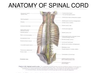
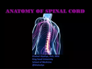
ANATOMY OF THE SPINAL CORD AND REFLEXES . PREPARED BY HUGH POTTER, Ph.D BIOLOGY DEPARTMENT UNION COUNTY COLLEGE. IMAGES USED WITH THE PERMISSION OF ADAM, INC. ANTERIOR VIEW OF THE SPINAL CORD. CERVICAL CORD (8 nerves).

E N D
ANATOMY OF THE SPINAL CORD AND REFLEXES PREPARED BY HUGH POTTER, Ph.DBIOLOGY DEPARTMENTUNION COUNTY COLLEGE IMAGES USED WITH THE PERMISSION OF ADAM, INC.
ANTERIOR VIEW OF THE SPINAL CORD CERVICAL CORD(8 nerves) The spinal cord begins at the base of the medulla oblongata and extends to about the 2nd lumbar vertebra. The cord is divided into four regions each of which has branches called spinal nerves. THORACIC CORD(12 nerves) LUMBAR CORD(5 nerves) SACRAL CORD(5 nerves)
SAGITAL SECTION OFLOWERSPINE The inferior, terminal portion of the spinal cord is at the level of the 2nd lumbar vertebra. Branches from the lumbar region pass downward from the cone-shaped tip (conus medullaris) of the spinal cord forming the cauda equina (horse’s tail). Cauda equina
The Meninges of the Spinal Cord The Meninges are tough, moist membranes surrounding the spinal cord. 1. Dura mater – The outermost membrane surrounding the spinal cord, it consists of a thin Meningeal layer. 2. Arachnoid layer – a fibrous, delicate middle zone. Below this layer is the subarachnoid space filled with cerebrospinal fluid. 3. Pia mater – innermost of the meningeal layers. It is closely adherent to the surface of the spinal cord.
SPINAL CORD CROSS SECTION A - ventral root of spinal nerveB - gray matterC - white matterD - dorsal root of spinal nerveE - spinal nerve D E B C A
CERVICAL PLEXUS The cervical plexus is formed by branches of cervical nerves C1 to C4 arising from the cervical region of the spinal cord. The phrenic nerve which stimulates the diaphragm to contract arises from the cervical plexus
The brachial plexus is formed from spinal nerves C5, C6, C7, C8 and T1. It is responsible for innervation of the skin and muscles of the entire upper limb and all but two muscles of the shoulder girdle. An accident that pulls the arm may damage the brachial plexus. In newborns, the brachial plexus can be damaged during birth if the delivery requires pulling the arm. BRACHIAL PLEXUS
LUMBAR PLEXUS Spinal nerves branching from the lumbar region of the cord form the lumbar plexus. Branches of this plexus stimulate muscles of the back, hip and thigh. The plexus also is responsible for sensation in the skin of the thighs, the pubic area and the external genitalia in males and females.
LUMBOSACRAL SPINAL CORDPOSTERIOR VIEW The area within the rectangle shows the lower portion of the spinal cord. The branches leaving the sacral region pass through the sacral foramina forming the sacral plexus.
SACRAL PLEXUS Spinal nerves branching from the lumbar (L4 and L5) and sacral (S1, S2, S3 and S4) region of the cord form the sacral plexus. Nerves branching from this plexus innervate the limb and pelvic area. Because the lumbar and sacral plexuses are interconnected, they are sometimes referred to as the lumbosacral plexus.
SCIATIC NERVE The sciatic nerve (astrerisk) is formed from spinal nerves arising from the lumbar (L4 and L5) and sacral (S1, S2 and S3) regions of the spinal cord. It passes into the thigh and lower leg supplying innervation of sensation and movement for the entire lower limb. When giving a gluteal intramuscular injection, it is important to inject into the gluteus medius muscle to avoid damage to this large nerve.
The Components of the Reflex Arc • 1. The sensory neurons enter the dorsal part of the spinal cord cell via the dorsal root of the spinal nerve. The cell bodies of these sensory neurons are located in a dorsal-root ganglionthat lies just outside the spinal cord near its dorsal side. • The axons of the sensory neurons then enter the spinal cord and synapse with interneurons within the gray matter of the spinal cord. • The interneurons in turn synapse with motor neurons, the axons of which exit the cord ventrally via the ventral root, and conduct information to the muscles
MONOSYNAPTIC REFLEX The patellar reflex is produced by striking the patellar tendon (arrow) with the reflex hammer. A stretch receptor in the tendon will send afferent impulses to the spinal cord. The incoming messages will synapse on motor neurons in the gray matter. These messages will travel out to the quadriceps muscle group (asterisk) producing the characteristic knee jerk. This type of reflex is called monosynaptic because the sensory neuron synapses directly on a motor neuron in the gray matter of the spinal cord.
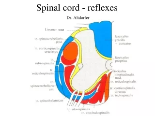
Major Spinal Nerve TractsMotor tracts – RedSensory tracts - Blue
Descending Nerve Tracts There are two major groups of descending tracts from the brain: the corticospinal, or pyramidal tracts, and the extrapyramidal tracts. The pyramidal tracts descend directly without synaptic interruption, from the cerebral cortex to the spinal cord. Pyramidal Tracts Corticobulbar tract Corticospinal tracts Extra-Pyramidal tracts Vestibulospinal tracts Tectospinal tracts
Descending tracts (Motor) Pyramidal Tracts – Originate from the pyramidal cells of the primary motor cortex. These nerve tracts decussate. Corticobulbar tract 1. Neurons originate in primary motor cortex of the cerebrum. 2. Destination is motor nuclei of cranial nerves in the brain stem. 3. Provide conscious control over skeletal muscles of the eye, jaw and face, as well as some muscles of the neck and throat. Corticospinal tracts 1. Neurons originate in primary motor cortex of the cerebrum. 2. Provide voluntary motor control of skeletal muscles throughout the body. 3. Destination of the tracts is the motor neurons of the ventral horns of gray matter in the spinal cord.
Extra-Pyramidal tracts – Originate from centers in the cerebrum, diencephalon and brain stem not from pyramidal cells (extra-pyramidal). Vestibulospinal tracts – do not decussate 1. Neurons respond to information from the vestibulocochlear nerve about the position and movements of the head. 2. The tract carries motor commands that alter muscle tone and position the head, neck and limbs to maintain balance and posture. Tectospinal tracts – Cross over in the brain stem 1. Neurons originate in the superior and inferior colliculi in the tectum of the midbrain. The colliculi receive visual (superior) and auditory (inferior) sensations. 2. Neurons of these tracts direct reflexive changes in the position of the head, neck and upper limbs in response to bright lights, sudden movements or loud noises.
Ascending Nerve Tracts The ascending tracts convey sensory information from cutaneous receptors, proprioceptors (muscle and joint senses), and visceral receptors. Most of the sensory information that originates in the right side of the body crosses over (decusses) and eventually reach the region on the left side of the brain, which analyses this information. Similarly, the information arising in the left side of the body is ultimately analysed by the right side of the brain. This decussation occurs in the medulla oblongata for sensory modalities, or in the spinal cord for other modalities of sensation. Ascending Tracts Posterior Column Pathway Spinothalamic Pathway Spinocerebellar Pathway
Ascending Tracts – Three major sensory pathways Posterior Column Pathway – Cross in the medulla oblongata Carries sensations of fine touch, pressure and proprioception from peripheral receptors to the sensory cortex of the cerebral hemispheres via the thalamus. Spinothalamic Pathway – Cross in the spinal cord Carries messages of crude touch, pressure, pain and temperature from peripheral receptors to the sensory cortex of the cerebral hemispheres via the thalamus. Spinocerebellar Pathway 1. Sensory neurons arise in muscle spindles, stretch receptors in tendons and joint capsules. The overall sensory information is proprioception. 2. The destination of these messages is the cerebellum, specifically the cerebellar cortex.
Autonomic Nervous System The nervous system is divided into the Somatic Nervous System which controls organs under voluntary control (mainly muscles) and the Autonomic Nervous System (ANS) which regulates individual organ function and homeostasis, and for the most part is not subject to voluntary control. It is also known as the visceral or automatic system. The ANS is predominantly an efferent (motor) system transmitting impulses from the Central Nervous System (CNS) to peripheral organ systems. Its effects include: 1. control of heart rate and force of heart contraction2. constriction and dilation of blood vessels3. contraction and relaxation of smooth muscle in various organs 4. visual accommodation and pupillary size 5. the secretions from exocrine and endocrine glands.
The Parasympathetic and Sympathetic Systems The ANS is divided into two separate divisions called the Parasympathetic and Sympathetic Systems, on the basis of anatomical and functional differences. Both of these systems consist of myelinated preganglionic fibers which make synaptic connections with unmyelinated postganglionic fibers, and it is these which then innervate the effector organ. These synapses usually occur in clusters called ganglia. Most organs are innervated by fibers from both divisions of the ANS, and the influence is usually opposing. For example, the vagus nerve (PNS) slows the heart, whilst the sympathetic nerves increase its rate and strength of contraction. In general, the responses of the parasympathetic system are characterized as “Rest and Digest” and those of the Sympathetic are called “ Fight or Flight”
Parasympathetic Nervous System Parasympathetic nerve messages arise from the cell bodies of the motor nuclei in the brain stem and from the second, third and fourth sacral segments of the spinal cord. For this reason, the parasympathetic is also referred to as Cranio-Sacral. Preganglionic fibers run almost to the organ which is innervated, and synapse in ganglia close to or within that organ, giving rise to postganglionic fibers which then innervate the relevant tissue.
Sympathetic Nervous System The cell bodies of the sympathetic preganglionic fibres are in the lateral horns of spinal segments T1 to L2. For this reason, the sympathetic nervous system is also called Thoraco-Lumbar. The preganglionic fibres travel a short distance in the mixed spinal nerve, and then branch off as white rami (myelinated) to enter the sympathetic ganglia. The sympathetic ganglia are mainly arranged in two chains which lie near the vertebral bodies and extend from the cervical to the sacral region. They are called the sympathetic ganglionic chains.
SYMPATHETIC CHAIN OF GANGLIA Running parallel on either side of the vertebral column is a chain of interconnected ganglia (arrow) that serve as relay stations for efferent sympathetic impulses that travel out to peripheral regions.
AUTONOMIC REFLEX The PUPILLARY REFLEX The pupillary reflex involves a change in the diameter of the pupil of the eye due to the interaction of the two branches of the autonomic nervous system. This response is usually elicited by shining a light into the eye stimulating the optic nerve (arrow). The autonomic nerves pass through cranial nerve III, the oculomotor nerve. The SNS will produce dilation of the pupil. The PNS will constrict the pupil.