Download
1 / 44
440 likes | 1.18k Views
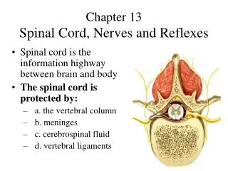
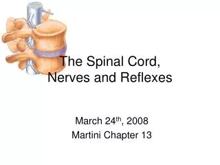
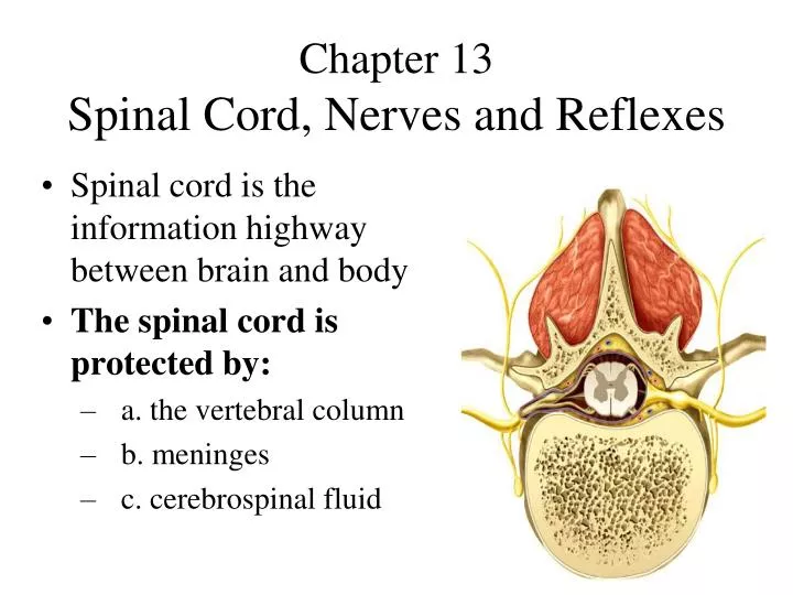
Chapter 13 Spinal Cord, Nerves and Reflexes. Spinal cord is the information highway between brain and body The spinal cord is protected by: a. the vertebral column b. meninges c. cerebrospinal fluid. Anatomy of the Spinal Cord.

E N D
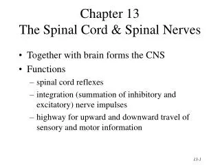
Chapter 13Spinal Cord, Nerves and Reflexes • Spinal cord is the information highway between brain and body • The spinal cord is protected by: • a. the vertebral column • b. meninges • c. cerebrospinal fluid
Anatomy of the Spinal Cord • 1. Spinal cord begins as a continuation of the medulla oblongata and terminates at about the L1 vertebra. • Thick as a finger • 2. Cervical and lumbar enlargements serve as points of origin for nerves to the extremities • 31 pairs of spinal nerves • 3. Distal end is tapered and called the cauda equina (horse’s_____)
The meninges are three fibrous coverings that enclose the spinal cord and brain a. innermost layer is the pia mater b. middle layer is the arachnoid c. outermost layer is the dura mater There is a “PAD” around the spinal cord Inflammation of the meninges is known as meningitis. Meninges of the Spinal Cord
Meninges of Vertebra & Spinal Cord • ____________ • ____________ • ____________
Spinal Tap/ Epidural • Placing a needle in the subarachnoid space is called a spinal tap (lumbar puncture) • Used to diagnose pathologies and to introduce drugs. • Headache is a frequent side effect, WHY? • Spinal tap must be given below L1, Why? • An Epidural is typically used for anesthesia
WHAT’S THE MATTER? • KINDS OF MATTER: • 1. White Matter- myelinated (Why white?) • 2. Gray Matter- unmyelinated (Why gray?) • 3. ______ (Alma Mater)
MORE ON MATTER • Spinal cord: • Gray matter forms an H-shaped inner core • Surrounded by white matter • Brain: • A thin outer shell of gray matter covers the cerebral hemispheres • THE SPINE IS GRAY INSIDE, WHITE OUTSIDE, THE BRAIN IS OPPOSITE
Cross-Sectional Anatomy of the Spinal Cord • Gray matter is shaped like the letter “H” or a butterfly and surrounded by white matter divided into 3 columns • Gray matter = neuron cell bodies • White matter = myelinated axons • Gray matter is divided into horns (posterior, anterior and lateral). • The posterior horn is the area of the posts
Gray Matter in the Spinal Cord • Pair of dorsal or posterior horns • totally sensory • Pair of ventral or anterior horns • totally motor fibers • Connected by gray commissure
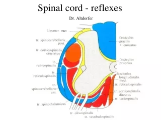
Spinal Tracts • Ascending tracts come from the periphery to head up and are called __________, _________ • Descending tracts head down and to the periphery are called ________, _________ • Decussation means that the fibers cross • Contralateral means origin and destination are on opposite sides while ipsilateral means on same side
The Motor and Sensory Brains The Grand prix (Pronounced: “grand pre”) is a motor car race
Sensory Homunculus • Demonstrates that the area of the cortex dedicated to the sensations of various body parts is proportional to how sensitive that part of the body is.
Motor Homunculus http://faculty.washington.edu/chudler/flash/fgames.html
Noisy Neighbors • Neurons that control your right arm and right leg dwell near each other in the _______ gyrus on the left side of your brain. • Experiment 1: Polish the desk with a clockwise circling motion of your right hand. Now start your right foot circling clockwise. • When neurons controlling your arm and leg are near each other they work well together. • Experiment 2: Now reverse the rotation direction of your right foot. • Tough to do, why? When neurons near each other are called on to do different work they disturb each other.
Neighbors Across Town • Your left arm and left leg are “across town” in the _____ precentral gyrus from your right and left arm and leg. • Experiment 3: Using you left arm to buff the top of your desk in a clockwise direction. This time rotate your right leg counterclockwise. • This should pose no problem because the control centers for the two limbs are found on opposite sides of the brain, so they don’t bother each other.
2 SENSORY TRACTS 1. Spinothalamic tract 2. Posterior (Dorsal) Columns These travel UP the spinal cord
Spinothalamic Tract • Pain and temperature • Decussation of the second order neuron occurs in spinal cord
Dorsal Columns • Vibration, proprioception • Two Dorsal columns on each side of the spine: • 1. Fasciculusgracile carry signals from the leg (remember: gracilis muscle of the leg) 2. Fasciculus cuneatus carry signals from the arm • Decussation of in medulla • Third neuron in thalamus carries signal to: ___ ___ of the brain
PROPRIOCEPTIONAwareness of your body in space ****** STAND UP ******
2 Motor Tracts Called the Pyramidal Tracts: 1. Anterior (Ventral) Corticospinal 2. Lateral Corticospinal These travel DOWN the spinal cord
Corticospinal Tract • Coordinates limb movements • Two neuron pathway start in the: • upper motor neuron in _____ _____ of the brain
The Spinal Nerves 31 pairs of spinal nerves: • a. 8 pairs of cervical nerves (C1-C8) • b. 12 pairs of thoracic nerves (T1-T12) • c. 5 pairs of lumbar nerves (L1-L5) • d. 5 pairs of sacral nerves, (S1-S5) • e. 1 pair of coccygeal nerves Spinal nerves exit superiorto their vertebrae for C1-C7 nerves only. C8 nerve-coccyx nerves exits inferior to their vertebrae
Spinal Nerves (Cont.) • Roots: points of attachment for each spinal nerve to a segment of the spinal cord • Posterior, or dorsal (sensory), root contains sensory nerve fibers, conducts nerve impulsesinto the spinal cord • The ganglion contains the cells bodies of the sensory neurons • Anterior, or ventral (motor), root contains motor neuron axons and conducts impulses fromthe cord • Cell bodies are located in the gray matter
Branches of a Spinal Nerve After passing through its intervertebral foramen (IVF), a spinal nerve divides into a dorsal and ventral branch called rami Spinal nerve branches, except for T2-T12, form a network of nerves called a plexus
Nerve Plexus • Two UP: • Cervical plexus, C1 to C5 • supplies neck and phrenic nerve to the diaphragm • Brachial plexus, C5 to C8 and T1 • supplies upper arm and shoulder, multiple nerves • Two DOWN: • Lumbar plexus, L1 to L4 • supplies anterior thigh & genitalia • Femoral nerve • Sacral plexus, L4- S4 • supplies butt & lower leg • Sciatic nerve
Cervical Plexus- Clinical • Damage to the cervical plexus can effect the diaphragm muscle. • Damage to the spinal cord in the origin of the phrenicnerves (C3-C5) causes respiratory arrest. • Breathing stops because the phrenic nerves no longer send impulses to the diaphragm. • Classic anatomy: “C3, 4 and 5 keeps the diaphragm alive.”
Brachial Plexus • Nerves of the upper extremities: • Axillary nerve to deltoid and teres minors and arm pit • Musculocutaneous nerve to flexors of arm and forearm and cutaneous sensation of forearm • Median nerve to anterior forearm, palm and first 3 ½ fingers (thumb, index finger, middle finger and lateral half of ring finger) • Ulnar nerve of anteriomedial forearm, palm and last 1 ½ fingers (medial half of ring finger, small finger) • Radial nerve to the posterior forearm and dorsal surface of hand
Brachial Plexus- Clinical • Injury to the brachial plexus: • Crutch palsy (Axillary nerves), • Wrist drop (Radial nerve) • Carpal tunnel syndrome (Median nerve) • Decreased sensation, thumb and wrist and pain • Tunnel of Guyon (Ulnar nerve) • decreased adduction, abduction of fingers, and flexing wrist, and sensation over little finger
Lumbar Plexus- Clinical • Injury to the femoral nerve • An inability to extend the leg • loss of sensation in the skin over the anteromedial aspect of the thigh.
Sacral Plexus- Clinical • The largest nerve in the body arises from the sacral plexus called the sciatic nerve. • A large as your small finger • Injury to the sciatic nerve results in pain from the buttock down the back of the leg, foot drop, an inability to dorsiflex the foot, and loss of sensation over the leg and foot. • Sciatica: Inflammation of the sciatic nerve • A few causes include: herniated (slipped) intervertebral disc, sacroiliac subluxation, piriformis syndrome
Dermatomes • Dermatome = skin sensation • All spinal nerves except C1 innervate segments of the skin • Helps diagnosis nerve problems • Shingles: acute infection of the peripheral nerves by the herpes zoster virus • The virus goes from the posterior horn and down the sensory nerves causing pain and skin blisters.
Reflex Arc • Simplest type of neural pathway • 1. receptor • 2. sensory neuron • 3. motor neuron • 4. effector • Example: The flexor (withdrawal) reflex is ipsilateral and is a protective withdrawal reflex that moves a limb to avoid pain • Brain is NOT involved in a reflex
The Muscle Spindle • Stretch receptors that monitor the length of skeletal muscles (biceps, triceps, petellar and achilles reflexes) • Golgi Tendon organs are stretch receptors in the tendon of a muscle.
Reflexes- Clinical • Reflexes are used to diagnose disorders and locating injured tissue. • Absent = usually not pathological • Absent unilaterally = damage may be somewhere along a nerve pathway on that side. • Hyperreflexia = Brain abnormality • Clinically important reflexes: • 1) Biceps (C5), Triceps (C7) • 2) Petellar (L4), Achilles reflex (S1) • 3) Babinski sign (Pathological reflex
Upper/ Lower Motor Neuron Disease • Paralysis • Upper motor neuron= spastic • Lower motor neuron= flaccid • UMN / LMN • UMN= Brain • LMN= peripheral nerves
CNS Nerve Injury • In the CNS, injury to the brain or spinal cord can be permanent. • Injury is followed by spinal shock • A loss of reflex activity called areflexia results. • Anti-inflammatory drugs given at time of trauma may decrease swelling • Spinal cord injury may result in paralysis which may be classified as: • monoplegia (one limb), diplegia (two limbs), paraplegia (both lower limbs)*, hemiplegia (one side), or quadriplegia (all limbs). • *Para = Beside; near; alongside
Polio and ALS • Diseases that cause destruction of motor neurons and result in skeletal muscle atrophy • Poliomyelitis is caused by poliovirus spread by contaminated water from fecal matter • weakness progresses to paralysis and possible respiratory arrest • Amyotrophic lateral sclerosis • sclerosis of spinal cord • paralysis and muscle atrophy
Spina Bifida Vera • Congenital defect • Failure of vertebral arch to close • Causes a lack of covering for the spinal cord • Mothers can reduce risk by taking folic acid supplement before pregnancy • Spina Bifida occultais a benign anamaly
Syphilis • Sexually transmitted disease • Three stages • Causes degeneration of the dorsal column • Symptoms will be loss of _______ and _______ (Dorsal column signs)