Download
1 / 30
300 likes | 543 Views
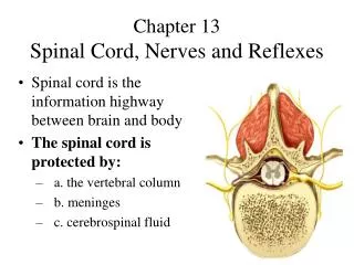
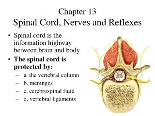
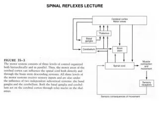
Spinal Nerves Reflexes. Objectives: Learn the structure of a spinal nerve Learn the branches of a spinal nerve Learn the four plexuses in the body and what they innervate Differentiate between intrinsic and acquired reflexes Learn the basics of a stretch reflex arc. Spinal Nerves Reflexes.

E N D
Objectives: Learn the structure of a spinal nerve Learn the branches of a spinal nerve Learn the four plexuses in the body and what they innervate Differentiate between intrinsic and acquired reflexes Learn the basics of a stretch reflex arc Spinal NervesReflexes
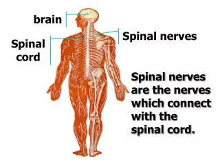
Gray Matter Ventral Horns Dorsal Horns Spinal Nerve Covered by perineurium Exits via IVF Spinal nerves have several branches Spinal Nerves
The two main branches of a spinal nerve are the... Dorsal Ramus Innervates the postural muscles of the back and the skin of the back Ventral Ramus Ventral rami form plexuses Spinal Nerves
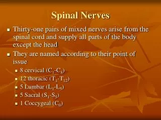
A Plexus is a complicated interlacing nerve network There are four plexuses in the body: The Cervical Plexus: C1-C4 The Brachial Plexus: C5-T1 The Lumbar Plexus: L1-L4 The Sacral Plexus: L4-S4 All plexuses give rise to named nerves. ONLY VENTRAL RAMI form plexuses Spinal Nerves
Hilton's Law: The nerves that innervate a joint also innervate the muscles that move the joint and supplies sensation to the skin around the joint. Spinal Nerves
Cervical Plexus: From the ventral rami of C1-C4 spinal nerve roots Superficial Branches: Lesser occipital (C2, C3) Greater Auricular (C2, C3) Trans. Cervical (C2, C3) Supraclavicular (C3, C4) Spinal Nerves
Cervical Plexus: From the ventral rami of C1-C4 spinal nerve roots Deep Branches: AnsaCervicalis (C1-C3) Phrenic Nerve (C3-C5) Spinal Nerves
Brachial Plexus Every anatomy student's nightmare. A complicated mess of nerves that arise from C5-T1 We'll break it down from proximal to distal: And no, you won't be responsible for every nerve! Spinal Nerves
Brachial Plexus: REAL (5) TEXANS(3) DRINK(6) COLD(3) BEER(5) Spinal Nerves
Brachial Plexus: Long Thoracic Nerve (C5-C7) Innervates Serratus Anterior Can be cut during mastectomy “Winged Scapula” Spinal Nerves
Brachial Plexus: Axillary Nerve (C5, C6): Innervates Deltoid, Teres Minor Can sometimes be damaged in childbirth ErbDuchenne's Palsy “Waiter's Tip” Spinal Nerves
Brachial Plexus: Musculocutaneous Nerve (C5-C7) Innervates the muscles of the Brachium Also implicated in ErbDuchenne's Palsy Spinal Nerves
Brachial Plexus: Median Nerve (C5-T1): Innervates muscles of the anterior forearm; flexors of the wrist and hand. Implicated in Carpal Tunnel and Pronator Teres syndrome Spinal Nerves
Brachial Plexus: Radial Nerve (C5-T1): Innervates the entire musculature of the posterior brachium and antebrachium All extensors of the elbow, wrist, and hand are innervated by the Radial nerve. Spinal Nerves
Brachial Plexus: Ulnar Nerve (C8, T1): Innervates the majority of the intrinsic muscles of the hand and thumb. “Funny Bone” Spinal Nerves
Lumbar Plexus (L1-L4) Responsible for the innervation of the superior aspect of the lower limb. Spinal Nerves
Iliohypogastric (L1): Skin of the upper abdomen Ilioinguinal (L1): Skin of the lower abdomen Genitofemoral (L1,L2): Cremasteric reflex in males, mons pubis and labiaemajora in females Femoral (L2, 3, 4): Hip flexors, knee extensors Obturator (L2, 3, 4): Adductors of the hip Spinal Nerves
Sacral Plexus: Responsible for innervation of the posterior and distal lower extremity. Innervates hip extensors and knee flexors, movers of the foot and ankle Spinal Nerves
Sciatic Nerve: L4-S3 Largest nerve in the body Doesn't directly innervate anything. Has no pain fibers “Sciatica” is actually nerve root pain and is often mis-diagnosed. Gives rise to two branches: Tibial and Common Fibular nn. Spinal Nerves
Tibial Nerve: L4-S3 Posterior leg muscles, plantar flexors of the foot and ankle. Skin of posterior leg, sole of foot Sensory supply to skin of posterior leg Spinal Nerves
Common Fibular: L4-S2 Has two branches; Superficial Fibular Innervates the muscles of the lateral cruris Responsible for eversion and plantar flexion of the foot Deep Fibular Innervates the muscles of the anterior cruris Responsible for Dorsiflexion of the foot Spinal Nerves
Dermatomes: An area of skin innervated by a single spinal nerve. Mapped out with great accuracy An excellent clinical tool to evaluate the level of injury in a patient Spinal Nerves
Motor endings and motor activity: The only things I want you to know... Acetylcholine is the neurotransmitter used to move muscles Acetylcholinesterase breaks down ACH, keeps the muscle from tetany Myasthenia Gravis Spinal Nerves
Reflex Arcs: Two general types of reflexes Intrinsic reflexes Learned reflexes All reflexes have a sensory component and motor component. Spinal Nerves
Reflex Components: Five components Receptor: Site of stimulus that starts the reflex Sensory Neuron: Carries the stimulus information to the spinal cord Integration Center: Where the sensory information gets transferred to the motor component of the reflex. Usually in the spinal cord. Reflexes will either be monosynaptic or polysynaptic. Simple reflexes are monosnaptic. Motor Neuron: Carries information from the integration center to the effector organ Effector: Muscle fiber or gland that responds to reflex impulses If reflexes act on skeletal muscle it's a somatic reflex Spinal Nerves
Stretch reflex: The Golgi Tendon Organs and Muscle Spindle Cells are responsible for this reflex The stretch reflex functions to keep us upright without our having to think about it As the Muscle Spindles are stretched, the associated muscle is stretched. This information is sent to the spinal cord to tell the associated skeletal muscle to contract. As the muscle contracts, the antagonist muscle relaxes (reciprocal inhibition) Spinal Nerves
Questions? Spinal Nerves