Download

1 / 120
1.22k likes | 1.25k Views
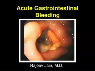
Gastrointestinal Bleeding. Case… . Hassan is 45 y/o saudi gentleman, presents to ED at KKUH early morning, C/O vomiting blood. How would you approach? How would you manage?. Gastrointestinal Bleeding. PERSPECTIVE Epidemiology

E N D
Case… • Hassan is 45 y/o saudi gentleman, presents to ED at KKUH early morning, C/O vomiting blood. • How would you approach? • How would you manage?
Gastrointestinal Bleeding PERSPECTIVE Epidemiology • Gastrointestinal (GI) bleeding is a relatively common problem countered in emergency medicine that often requires early consultation and hospital admission.
Gastrointestinal Bleeding • The overall mortality rate for GI bleeding is approximately 10% and has not changed significantly since the 1960s. • Diagnostic modalities have improved much more than therapeutic techniques.
Gastrointestinal Bleeding • GI bleeding is often easy to identify when there is clear evidence of vomiting blood or passing blood in the stool, but the clinical presentation may be subtle, with signs and symptoms of hypovolemia, such as dizziness, weakness, or syncope.
Gastrointestinal Bleeding • The approach to GI bleeding depends on whether the hemorrhage is located in the proximal or the distal segment of the GI tract (i.e., upper or lower GI bleeding). • These segments are anatomically defined by the ligament of Treitz in the duodenum.
Gastrointestinal Bleeding • Lower GI bleeding (LGIB) affects a smaller portion of patients and results in proportionally fewer hospital admissions than UGIB.
Gastrointestinal Bleeding • GI bleeding can occur in persons of any age but most commonly affects people in their 40s through 70s (mean age, 59 years). • Most deaths caused by GI bleeding occur in patients older than 60 years. UGIB is more common in men than in women (in a 2 : 1 ratio), whereas LGIB is more common in women.
Gastrointestinal Bleeding • Significant UGIB requiring admission is more common in adults, whereas LGIB requiring admission is more common in children.
Gastrointestinal Bleeding DIAGNOSTIC APPROACH Differential Considerations • Peptic ulcer disease, gastric erosions, and varices account for approximately three fourths of adult patients with UGIB.
Gastrointestinal Bleeding • Diverticulosis and angiodysplasia account for approximately 80% of adults with LGIB. In children, esophagitis, gastritis, and peptic ulcer disease are the most common causes of UGIB, and infectious colitis and inflammatory bowel disease are the most common causes of LGIB.
Gastrointestinal Bleeding • In children younger than 2 years of age, massive LGIB is most often a result of Meckel’s diverticulum or intussusception. • At all ages, anorectal abnormalities are the most common cause of minor LGIB.
Gastrointestinal Bleeding • Despite improved diagnostic techniques, no source of bleeding is identified in approximately 10% of patients with GI bleeding. • In patients with abdominal aortic grafts who present to the emergency department (ED) with GI bleeding, the possibility of aortoenteric fistula should be considered.
Gastrointestinal Bleeding • Prompt surgical consultation in the ED should be obtained if this is suspected, because bleeding can be massive and fatal.
Gastrointestinal Bleeding Rapid Assessment and Stabilization • Most patients with GI bleeding are easy to diagnose because they present to the ED complaining of vomiting blood or passing black or bloody stool. • The diagnosis is confirmed quickly by examination of the stool for the presence of blood.
Gastrointestinal Bleeding • Patients with suspected GI bleeding who are hemodynamically unstable should undergo rapid evaluation and resuscitation. • They should be undressed quickly to permit placement of cardiac and oxygen saturation monitors, and supplemental oxygen should be given as needed.
Gastrointestinal Bleeding • At least two large-bore peripheral intravenous lines should be placed (minimum 18-gauge); blood should be drawn for hemoglobin or hematocrit, platelet count, prothrombin time (PT), and type and screen or type and crossmatch studies; and crystalloid resuscitation should be initiated.
Gastrointestinal Bleeding • Intravenous crystalloid fluid should be given as a 2-L bolus in adults or 20 mL/kg in children until the patient’s vital signs have stabilized or the patient has received 40 mL/kg of crystalloid in an adult or 60 mL/kg as a child.
Gastrointestinal Bleeding • Patients who remain unstable after 40 to 60 mL/kg of crystalloid should be given type O, type-specific, or cross matched blood, depending on availability. • Persistently unstable patients should receive immediate consultation with a gastroenterologist for UGIB and with a surgeon for LGIB.
Gastrointestinal Bleeding • History, physical examination, testing a stool sample for blood, and measuring hemoglobin or hematocrit are the keys to diagnosing GI bleeding in most patients.
Gastrointestinal Bleeding History • Patients typically complain of vomiting red blood or coffee grounds–like material, or passing black or bloody stool. • Hematemesis (vomiting blood) occurs with bleeding of the esophagus, stomach, or proximal small bowel.
Gastrointestinal Bleeding History • Approximately 50% of patients with UGIB present with this complaint. • Hematemesis may be bright red or darker (i.e., coffee grounds–like) as a result of the conversion of hemoglobin to hematin or other pigments by hydrochloric acid in the stomach.
Gastrointestinal Bleeding • The color of vomited or aspirated blood from the stomach does not differentiate between arterial and venous bleeding. • Melena, or black tarry stool, will result from the presence of approximately 150 to 200 mL of blood in the GI tract for a prolonged period.
Gastrointestinal Bleeding • Melena is seen in approximately 70% of patients with UGIB and in one third of patients with LGIB. • Black stool that is not tarlike may result from presence of 60 mL of blood from the upper GI tract. Blood from the duodenum or jejunum must remain in the GI tract for approximately 8 hours before turning black.
Gastrointestinal Bleeding • Occasionally, black stool may follow bleeding into the lower portion of the small bowel and ascending colon. • Stool may remain black and tarry for several days, even though bleeding has stopped.
Gastrointestinal Bleeding • Hematochezia, or bloody stool (bright red or maroon), most often signifies LGIB but may be due to a brisk UGIB with rapid transit time through the bowel in 10 to 15% of patients. • Because UGIB is much more common than LGIB, a more proximal source of significant bleeding must be excluded before assuming the bleeding is from the lower GI tract.
Gastrointestinal Bleeding • Approximately two thirds of patients with LGIB present with red blood from bleeding per rectum. • Small amounts of red blood (e.g., 5 mL) from rectal bleeding, such as bleeding due to hemorrhoids, may cause the water in the toilet bowl to appear bright red.
Gastrointestinal Bleeding • Bright red stools also can be seen after ingestion of a large quantity of beets; in this case, Hemoccult testing would be negative and the patient also will report pink colored water in the toilet bowl.
Gastrointestinal Bleeding • In taking the history, specific questions should address the duration and quantity of bleeding, associated symptoms, previous history of bleeding, current medications, alcohol, nonsteroidal anti-inflammatory drug use and long-term aspirin ingestion, allergies, associated medical illnesses, previous surgery, treatment by nonhospital personnel, and the response to that treatment.
Gastrointestinal Bleeding • Patients with GI bleeding may report symptoms of hypovolemia, such as dizziness, weakness, or loss of consciousness, most often after standing up. • Other nonspecific complaints include dyspnea, confusion, and abdominal pain.
Gastrointestinal Bleeding • Rarely an elderly patient may present with ischemic chest pain precipitated by significant anemia due to a GI bleed. • One in five patients with GI bleeding may have only nonspecific complaints.
Gastrointestinal Bleeding • The history is of limited help in predicting the site or quantity of bleeding. • Patients with a previously documented GI lesion bleed from the same site in only 60% of cases.
Gastrointestinal Bleeding • Gross estimates of blood loss based on the volume and color of the vomitus or stool (e.g., brown or black, pink or red) or the number of episodes of hemorrhage are notoriously inaccurate.
Gastrointestinal Bleeding Physical Examination • VitalSigns Vital signs and postural changes in heart rate and blood pressure have been used to assess the amount of blood loss in patients with GI bleeding but are insensitive and nonspecific, with the exception of significant, sustained heart rate increase and hypotension.
Gastrointestinal Bleeding • All patients with a history suggesting GI bleeding who are hypotensive, are tachycardic, or experience sustained posture-induced changes in heart rate of greater than 20 beats per minute should be assumed to have a significant hemorrhage.
Gastrointestinal Bleeding • Normal vital signs do not exclude a significant hemorrhage, and postural changes in heart rate and blood pressure may occur in individuals who are not bleeding (e.g., elderly patients, many normal individuals, individuals on certain medications such as beta-blockers, individuals with hypovolemia from other causes).
Gastrointestinal Bleeding • General Examination The physical examination is valuable in establishing a specific diagnosis and assessing the severity of blood loss and the physiologic response to that loss.
Gastrointestinal Bleeding • Careful attention is given to the patient’s general appearance, vital signs, mental status (including restlessness), skin signs (e.g., color, warmth, and moisture to assess for shock, or presence of lesions such as telangiectasia, bruises, or petechiae to assess for vascular diseases or hypocoagulable states), pulmonary and cardiac findings, abdominal examination, and rectal and stool examination.
Gastrointestinal Bleeding • Frequent reassessment is important because a patient’s status may change quickly.
Gastrointestinal Bleeding • Rectal Examination Rectal and stool examinations are often key to making or confirming the diagnosis of GI bleeding. • The finding of red, black, or melenic stool early in the assessment is helpful in prompting early recognition and management of patients with GI bleeding.
Gastrointestinal Bleeding • The absence of black or bloody stool, however, does not exclude the diagnosis of GI bleeding. • Regardless of the apparent character and color of the stool, occult blood testing is indicated.
Gastrointestinal Bleeding Ancillary Testing • Tests for Occult Blood The presence of hemoglobin in occult amounts in stool is confirmed by tests such as ( Hemoccult, HemaPrompt). • Stool tests for occult blood may have positive results 14 days after a single, major episode of UGIB.
Gastrointestinal Bleeding • False-positive results have been associated with the ingestion of certain fruits (e.g., cantaloupe, grapefruit, figs), uncooked vegetables (e.g., radish, cauliflower, broccoli) and red meat, methylene blue, chlorophyll, iodide, cupric sulfate, and bromide preparations.
Gastrointestinal Bleeding • False-negative results are uncommon but can be caused by bile or ingestion of magnesium containing antacids or ascorbic acid. • Tests to evaluate gastric contents for occult blood (e.g., Gastroccult) can be unreliable and should not be used for this purpose. • In newborns, maternal blood that is swallowed may cause bloody stools; the Apt test may show that it is maternal in origin.
Gastrointestinal Bleeding • Clinical Laboratory Tests Blood should be drawn for evaluation of baseline hematocrit or hemoglobin, coagulation studies (PT and platelet count), and type and crossmatch studies (or type and screen studies if the patient is stable). • The initial hematocrit may be misleading in patients with preexisting anemia or polycythemia.
Gastrointestinal Bleeding • Changes in the hematocrit may lag significantly behind actual blood loss. Infusion of normal saline speeds equilibration of the hematocrit; however, rapid infusion of crystalloid in nonbleeding patients also may cause a decrease in hematocrit by hemodilution.