Download
1 / 50
1.17k likes | 4.6k Views
Gastrointestinal Bleeding . Chase R. Herdman , MD. Gastroenterology Associates of North Texas. Gastrointestinal Bleeding. Definitions and Classification of GIB UGIB Etiology LGIB Etiology Initial Assessment and Management Diagnosis Endoscopic Treatment Conclusions. GI Bleeding.

E N D
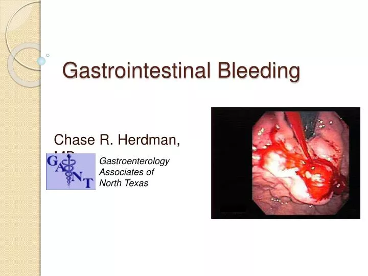
Gastrointestinal Bleeding Chase R. Herdman, MD Gastroenterology Associates of North Texas
Gastrointestinal Bleeding • Definitions and Classification of GIB • UGIB • Etiology • LGIB • Etiology • Initial Assessment and Management • Diagnosis • Endoscopic Treatment • Conclusions
GIBleeding • Common and potentially life threatening condition, which results in hemodynamic instability, anemia, or the need for blood transfusion. • Occult GI bleed • Blood in the feces too small to be seen but detectable by chemical tests
Ligament of Treitz • Ligament of Treitz: a fibrous band by which the duodenojejunal junction is fixed to posterior wall of abdominal cavity • Upper GIB: Bleeding arising proximal to LOT • Lower GIB: Bleeding arising distal to LOT
Definitions • Hematemesis • Vomitus of red blood or "coffee-grounds" material. • Represents an upper GI bleed. • Hematochezia • The passage of bright red or maroon blood from the rectum. • Represents lower GI bleeds (90%) or severe upper GI bleeds (10%) • Melena • Black, tarry, foul-smelling stool resulting from digested blood (at least 100 ml) • Usually indicates upper GI bleed, but can represent small bowel or right-sided colonic hemorrhages if an obstruction is present or if transit time is otherwise prolonged • Estimated to take approximately 8 hours to turn stool black
Black Stool • Non-GIB causes of black stool include: • Bismuth subsalicylate (Pepto-Bismol®) • Iron • Spinach • Charcoal • Dark beers (stout or Guinness) • Swallowed blood from nose bleeding
Hematemesis Hematochezia Melena PUD Esophageal varices Mallory-Weiss tear Esophagitis Vascular Anomalies (Telangiectasia, Angiodysplasia) gastritis Erosive Esophagitis Gastric Neoplasms (carcinoma, lymphoma, leiomyoma, sarcoma) Esophageal cancer Diverticular disease Angiodysplasia Colorectal carcinoma IBD Hemorrhoid Anal fissure Colonic polyp Brisk upper GI or small bowel bleed Meckel’s diverticulum Crohn’s disease All causes of hematemesis Neoplasm (rare) Another Classification of GIB
Upper Gastrointestinal Bleeding • Over 350,000 US hospitalizations/yr • Cost $1 billion/yr • Mortality rate: 10%; more common males • Rarely die from exsanguinations but rather from complication of an underlying disease • Self-limited in 80% of pts
UGIB - Etiology • Peptic Ulcer Disease (20-55%) GU > DU • Esophagogastric varices (14%) • Mallory-Weiss Tears (5%) • Vascular anomalies (6%) • Hemorrhagic and erosive gastropathy or gastritis (3-11%) • Erosive Esophagitis (2-8%) • Gastric Neoplasms (1-4%) • Others (7-25%) • Aortoenteric fistula, hemobilia, pancreatic malignancy
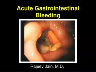
Peptic Ulcer Disease • Half of major UGIB w/ mortality of 6-10%. • Chronic ulcers, caused by exposure to acid & pepsin • Usually solitary • Size ranges from ~ 0.6 - 4 cm • Most common in duodenum and antrum
Peptic Ulcer Disease • Clinical: epigastric pain 1-3 hours after meals; nausea; vomiting; weight loss, acute onset bleeding • Complications: • Hemorrhage - 25% of ulcer deaths • Perforation – 66% of ulcer deaths • Obstruction • Malignant transformation
Risk Factors for peptic ulcers • H. pylori • NSAIDS • Stress • Gastric Acid
H. Pylori • Transmitted fecal-oral • Disrupts mucous layer, liberating enzymes and toxins causing mucosal damage • Assoc. w/ gastric cancer and non-Hodgkin’s gastric lymphoma • Eradication should be sought for recurrence prevention
NSAIDS • Inhibition of prostaglandins • Most of these ulcers are asymptomatic and uncomplicated • Implicated as an important factor for non-healing ulcers
Stress related ulcers • Generally refers to physiologic stress • Increased risk in hospitalized patients with life threatening non-bleeding illness • Increased risk with respiratory failure or coagulopathy • Ulcer prophylaxis with daily H2 blockers or PPI therapy • Some evidence to support psychologic stress may also be a cause
Gastric Acid • Rarely is hyperacidity the only contributor to peptic ulcer formation • Usually coupled with H. pylori, NSAIDS, stress which leads to increased permeability to back diffusion of hydrogen ions resulting in ulceration • Zollinger-Ellison syndrome
Portal Hypertension • Bleeding from varices: • esophageal • Less common gastric or duodenal • Usually stops spontaneously 50% • Mortality rate near 70-80% for those with continued bleeding • Mortality risk 30% with each recurrent bleed • Untreated 50% will rebleed during hospitalization • Prophylaxis: propranolol can decrease risk of rebleeding vs. endoscopic variceal ligation for high risk patients
Mallory-Weiss Tears • Tear usually induced by vomiting, often seen in Etohism • Tearing occurs when the cardia has been forced into the thorax • Morphology: irregular longitudinal tear in the esophago-gastric junction. May involve only the mucosa, or may rarely penetrate the wall. • 5-10% of UGI bleeding • When a Mallory-Weiss tear penetrates all layers of the wall, it is called Boerhaave’s syndrome. • Boerhaave’s tears may cause mediastinitis or peritonitis.
Dieulafoy’s lesion • Uncommon cause of major GIB. • arteriole that protrudes through a tiny mucosal defect causing a single small ulcer • usually within 6 cm of the gastroesophageal junction on the lesser curvature • Can occur in distal esophagus, small intestine, colon, and rectum.
Vascular Anomalies • Found throughout the GI tract; chronic or acute bleeding • 7% of UGIB. • Examples: • Vascular ectasias (angiodysplasias) • bright red stellate appearance. • Hereditary hemorrhagic telangiectasia (Osler-Weber-Rendu) • gastric antral vascular ectasia (GAVE or "watermelon stomach“) • CREST syndrome • Incidence increased in chronic renal failure
Gastritis • endoscopically visualized subepithelial hemorrhages and erosions • mucosal lesions, thus do not cause major bleeding. • Rarely causes UGIB (5%) • more commonly results in chronic blood loss • Common causes: • ingestion of NSAIDs • 50% of patients who chronically ingest NSAIDs. • Alcohol • 20% of actively drinking alcoholic patients • stress (severe medical or surgical illness).
Erosive Esophagitis • Severe erosive esophagitis due to chronic GERD may rarely cause UGIB. • Other Predisposing Factors: • Infections (esp. in immunosuppressed patients- candida, CMV, HSV) • Ingestion of irritants: alcohol, peppermint, tomato products, smoking, caffeine • Uremia • Cancer chemotherapy, radiation therapy • Prolonged NG tube
Lower Gastrointestinal Bleeding • Hospital mortality < 3%. Common in adult women and children. • 200-fold increase in incidence 3rd to 9th decade • Spontaneous cessation of bleeding > 85%. • Less likely to present in shock; require less transfusions
Etiology • Etiology varies with age • Elderly – diverticulosis, angiodysplasia, neoplasms, or ischemia • Young adults – infectious colitis, anorectal disease, inflammatory bowel disease, diverticulosis, or angiodysplasia • Children – Meckel’s diverticulum or intussusception • No source identified in ~20% of cases
Diverticulosis • Diverticula • Outpouchings in the colon • Thin wall, lined by mucosa & submucosa but no muscularispropria. • As it herniates, the vasa recta are exposed to injury on the luminal side leading to intimal thickening and media thinning which predisposes to rupture • Found in half of people over the age of 50, but <15% of these patients develop LGIB. • The most common cause of major LGIB (50% of cases). • Pathogenesis: low fiber diet stool bulk peristaltic contractions intraluminal pressure diverticula.
Diverticulosis • Diverticula are more prevalent in the left colon, but bleeding occurs more in the right. • S&S’s: acute, painless, large-volume maroon or bright red hematochezia in pt over age 50. • Subsides spontaneously in 80% of people but may recur in up to 25%. • > 95% of cases requires < 4 units of blood transfusion; 15-25% require surgery • Mortality is 5-20%.
Angiodysplasia/AV Malformations • Dilated torturous submucosal vessels • Causes painless bleeding ranging from melena or acute hematochezia to chronic occult blood loss. • 5-10% of LGIB • Occur commonly in the distal ileum, cecum and ascending colon. • Bleeding usually episodic and self limited • Re-bleeding occurs in up to 85% who are untreated • Generally treated endoscopically
Ischemic Colitis • More common in elderly • Mostly those with known atherosclerosis • Other causes: hypotension, CHF, arrhythmia • Watershed areas such as the splenic flexure and rectosigmoid junction.
Ischemic Colitis • 5% of pts after surgery for ileoaortic or abdominal aortic aneurysm. • Young pts may develop colonic ischemia due to vasculitis, coagulation disorders, estrogen therapy, and long distance running. • Presents as hematochezia and/or bloody diarrhea typically associated with mild cramps. • Bleeding is generally mild and self-limited. • Appearance can be similar to IBD, so clinical history is important
Inflammatory Bowel Diseases • Pt w/ Crohn’s Disease or Ulcerative Colitis often have diarrhea with hematochezia, fever, dehydration. • UC a more common cause for bleeding than Crohn’s • Associated with urgency, tenesmus, abdominal pain/cramping. • Rarely causes massive bleeding, but if it does => surgical intervention.
Neoplasms • Benign polyps (ulcerated) and carcinoma: chronic occult blood loss or intermittent anorectal hematochezia. • Colonic neoplasms cause 10% of acute LGIB. • Benign Polyps • Adenomatous: Tubular, Tubulovillous,Villous variants • 90% of APs in the colon, progress in >10 yrs to adenocarcinoma • Hamartomatous (Peutz-Jeghers) • Malignant: • Adenocarcinoma • 98% of all cancer of the colon; peak age is 60-79 yr for sporadic cases • predisposing factors include adenomatous polyps, UC, genetic factors (defective APC tumor suppressor gene), low fiber, high animal fat diet
Other Causes • Radiation-induced proctitis/colitis. • Usually occurs years later, but can occur any time following radiation therapy • Aortoenteric fistula in patients with prior history of aortic surgery. • Vasculitis syndromes. • NSAID induced ulcers • Post-polypectomy bleeding • Post-sphincterotomy bleeding • Infectious colitis
Assessment • HPI • Characteristics of bleeding, onset and duration of bleeding, associated symptoms, etc. • Medications • Aspirin, NSAIDs, warfarin, Plavix, Beta blockers, anti-HTN meds • PMH • H/o dyspepsia or PUD suggests peptic ulcer; H/o of vomiting, retching, or coughing preceding hematemesis in an alcoholic pt suggests Mallory-Weiss tear • FH • Liver disease, bleeding disorders, Osler Weber Rendu • Physical Exam • VS (orthostatic?) • Tachycardia, BP, O2 sats • Signs of chronic liver disease • Jaundice, ascites, edema, spider angiomata implicate bleeding due to portal hypertension • Vascular status • Capillary refill, peripheral pulses • Abdominal exam • Rectal exam • Labs: • CBC, PT, PTT, INR, BUN, serum creatinine, LFT, and cross-matching for 2-4 units or more of PRBC • Note: UGIB is suggested by a BUN/Cr ratio > 30:1 • this is due to digested blood in bowel and prerenalhypovolemia • Imaging studies – r/o perforation
Assessment & Initial Management • Initial Triage: • Very Low Risk: Normal hemodynamics, no overt bleeding within 48 hrs, neg NG lavage, normal lab tests, and no serious comorbid illnesses or advanced liver disease • Does not require hospital admission. • Low to moderate risk • Admit and undergo endoscopy usually within 24 hrs. • High risk: Active bleeding (hematemesis or bright red blood on NG aspirate), loss of > 5 units of blood, persistent hemodynamic derangement despite fluid resuscitation, serious comorbid med illness, or advanced liver disease • ICU admission. • GI +/- surgical consult • Intubation?
Initial Management • ABC’s (no “E”) in initial management • Fluid resuscitation and Blood Replacement: • 2 large bore IVs or central line • PRBC given to maintain a hematocrit of 25-30% • Platelets if < 50,000/ul or if there is impaired platelet function due to aspirin/clopidogrel use • FFP for actively bleeding pts with coagulopathy and INR>1.5 • Discontinue aspirin, NSAIDs, and anticoagulants. • NG Tube • If actively vomiting • If suspected UGIB • Aspiration of red blood or “coffee grounds” confirms an UGIB. • 10% have nonbloody aspirates (doesn’t rule out UGIB)
Acute Pharmacologic Therapies • Acid inhibitory therapy: • IV PPI • Omeprazole or pantoprazole, 80 mg bolus, followed by 8mg/hr continuous infusion for 72hrs • Reduces the risk of rebleeding in pts w/ peptic ulcers w/ high risk features after endoscopic therapy. • Active bleeding, visible vessel, adherent clot • High doses of oral PPI • As effective as IV but slower time to pH >6. • Octreotide: • IV 100ug bolus, followed by 50-100ug/h • Reduces splanchnic blood flow and portal blood pressures • Effective initial control of bleeding related to portal HTN • Emperic use in suspected UGIB with evidence of liver disease or known portal HTN until the source of bleeding can be determined by endoscopy
Diagnosis • Upper Endoscopy • Preferably when hemodynamically stable • Continued active bleeding may require more urgent endoscopic evaluation • Consider intubation • Colonoscopy depending on clinical history or lack of findings on EGD • Small bowel evaluation • Imaging • Angiography
Diagnosis • Small Intestine Push Enteroscopy: • To evaluate bleeding from small bowel (ie. Vascular ectasias). • Consists of passage of a long, small diameter endoscope that may reach from the proximal to the distal jejunum. • Capsule Imaging: • a wireless video capsule device ingested and travels the SB as video images are transmitted to a portable recorder. • Allows to identify vascular ectasias, neoplasms and other bleeding lesions but does not permit precise localization or therapy. • Not always available as inpatient
Diagnosis • Nuclear Bleeding Scan (Technetium-99m labeled RBC): • Can localize slow bleeding (0.1ml/min) to the SI, right colon, or left colon. • Accuracy of a positive study is only 78% b/c the bleeding may be slow or intermittent. • Low risk (less invasive) • Usually used as a preliminary study to target confirmatory studies (angiography or colonoscopy). • Angiography (Selective Mesenteric Angiography): • Used more for LGIB than UGIB • Localizes source of bleeding at a rate of 0.5 ml/min or greater. • Accurate 50-75% of time in patients who are actively bleeding. • Complication rate is 3%: stroke, renal failure, and thrombosis. • Allows for possible intravascular treatment of bleeding lesion • Intra-Arterial Vasopressin • Intra-arterial embolization/coiling
Endoscopic Management • Variceal Bleeding • Esophageal banding • Injection therapy • Non-Variceal Bleeding • Injection therapy • Contact thermal probes • Hemoclip • APC
Endoscopic Treatment of Esophageal Varices • Octreotide IV • Antibiotics • Esophageal banding • Often used as secondary prophylaxis • Sclerosant injection • 5% Sodium morrhuate • 5% Ethanolamine • At least one of above should be readily available • Isolated gastric or duodenal varices not amenable to above therapies • Cyanoacrylate glue • TIPS • Surgery
Endoscopic Therapy of Nonvariceal Bleeding • Injection therapy • Epinephrine 1:10,000 mix • Used in combination with another modality • Contact thermal probes • Multipolar (eg, Gold probe) +/- inj needle • Firm taponade • 12-14 W • 7-10 seconds • Heater probe • 4-5 pulses • 30 joules • Complete flattening of vessel is goal
Endoclip placementOlympus QuickClip, Boston Scientific Resolution Clip, Wilson Cook TriClip, InScope multiclip • Pros and Uses • Generally considered as effective as thermal modalities • Often combined with injection therapy of epinephrine • Must be placed firmly against tissue • Preferably perpendicular to vessel anatomy • Used to control bleeding • Small ulcers with visible vessel • Dieulafoy lesions • Mallory Weiss tears • Diverticular bleeding • AVMs • Cons and Limitations • Large ulcers • Cardia lesions • Tight spaces • Orientation • Limited supply • Other uses • Closure of perforation, fistula, EMR defects • Angiographic marker • Feeding tube retention
APC (argon plasma coagulation) • Argon gas combined with high voltage spark from catheter • Ionizes gas delivering energy to tissue in close proximity • 2-3 mm depth • Does not require tissue contact • 20-40 W depending on location • Uses • AVMs • GAVE • Radiation telengectasias • Piecemeal polypectomy • Ulcers • Polyp/Tumor ablation • Cons and complications • Availability • Bowel insufflation • Tissue contact
Other Treatment • Transvenousintrahepaticportosystemic shunts (TIPS): • Performed percutaneously through the jugular vein and involves the creation of a permanent intrahepatic shunt between the hepatic and portal veins, easing portal hypertension. • Indicated in pts in whom endoscopy has failed to control acute variceal bleeding. • TIPS decreases rebleeding more effectively than endoscopic therapy. • Surgery • Indications: • All hemodynamically unstable patients with active bleeding who do not respond to intravascular volume replacement and correction. • Active bleeding that requires > 4-6 u blood within 24h or > 10 total units. • Preoperative localization of the bleeding site by endoscopy, nuclear imaging or angiography => allows limited resection of the bleeding segment of SB and colon. • If accurate localization is not possible or if emergency surgery is required for massive bleeding => total abdcolectomy with ileorectalanastomosis => increases morbidity and mortality more than limited resections. • Re-bleeding rate is less than 10%.
Conclusions • GI bleeding a common cause for hospitalization and mortality • History provides many keys to location of bleeding • Resuscitation, Resuscitation, Resuscitation • Equipment review and on cart • Eg, bleeding buckets • Boating analogy • Anticipate equipment needs during endoscopy