Download
1 / 51
650 likes | 1.33k Views
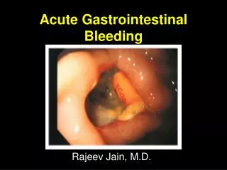
Gastrointestinal Bleeding. Asaf Kedar M.D. Department of Surgery Hadassah-Hebrew University Medical Center. Introduction. 1-2% of acute hospital admissions. Leading diagnosis in patients admitted to ICU. Mortality overall – 5-12%, with persistent or recurring hemorrhage – 40%.

E N D
Gastrointestinal Bleeding AsafKedar M.D. Department of Surgery Hadassah-Hebrew University Medical Center
Introduction • 1-2% of acute hospital admissions. • Leading diagnosis in patients admitted to ICU. • Mortality overall – 5-12%, with persistent or recurring hemorrhage – 40%. • Up to 85% cease spontaneously. • Hemorrhage can arise in any part of the GI track from nose / mouth to anus, including organs that empty into the GI.
Introduction • Upper Gastrointestinal Bleeding (UGIB) – Proximal to the ligament of Treitz. • Lower Gastrointestinal Bleeding (LGIB) – Distal to the ligament of Treitz. • >85% of major bleeding are d/t: • PUD • Variceal hemorrhage • Colonic diverticulosis • Angiodysplasia. Aorta Cisterna Chyli Celiac Ganglia Ligament of Treitz Portal vain Jejunum Duodenum Superior Mesenteric vessels
Introduction • Only age is a risk factor for hemorrhage • Surgery is required in 5-10% of patients hospitalized with GI bleeding. • Four primary goals in initial management of a patient with acute GIB: • Patient assessment – Hemodynamic status and identification of significant comorbidities. • Resuscitation and monitoring. • Identification of the source of bleeding. • Specific therapy
Initial Patient Assessment • Characteristics of bleeding: • Hematemesis • Melena • Hematochezia • Associated symptoms: • dizziness, syncope, antecedent dyspepsia, crampy abdominal pain, wt. loss. • Medications: • Salicylates, NSAIDs, clopidogrel (plavix), warfarin, clexan, ß blockers, Ca-channel blockers, anti-hypertensives. • Past Medical History: • GIB, dysphagia, GERD, H.pylori infection, PUD, liver disease, alcohol abuse, IBD, diverticulosis, malignancy.
Initial Patient Assessment • Co-morbid medical conditions: • Renal insufficiency, IHD, CHF, chronic respiratory disease, liver disease, CNS disability. • Physical examination: • Determine the degree of blood loss, • Rectal examination • exclude nasopharyngeal bleeding. • Assesment of cirrhosis – Jaundice, ascitis, palmarerythema, caput medusa.
Initial Patient Assessment • Melanin spots on lips, oral mucosa and digits, small intestinal polyposis Peutz – Jeghers syndrome. • Cutaneoustelangiectasias. Osler – Weber – Rendu Syndrome
Initial Patient Assessment • Initial Laboratory Assessment: • Biochemistry profile • CBC • Coagulation profile • Type and cross-matching • HGb less than 10g/100ml is associated with increased risk for morbidity and mortality.
Resuscitation • 2 large-bore IV lines • Crystalloids • Colloids • Packed cells and other blood products • Foley catheter • Central venous or pulmonary artery catheter. • Endotracheal intubation for massive hematemesis and mental obtundation, hemodynamically unstable patient. • Patient with GIB rarely die from hemorrhage – they die from multisystem organ failure d/t shock Treat fast to prevent and reverse these processes ASAP
Identification of Source of Bleeding • Nasogastric tube (NGT) of large caliber.
Identification of Source of Bleeding • Patients with suspected upper GIB require upper endoscopy with diagnostic and therapeutic capabilities. • Timing of examination – from immediately to within 24h for stable patients.
Identification of Source of Bleeding • Patients with suspected lower GIB require colonoscopy with diagnostic and therapeutic capabilities (after colonic purging). • Hemodynamically stable patient with hematochezia / melena with negative upper gastrointestinal examination may be presumed to have acute lower GIB, choice of initial diagnostic test remain controversial.
Identification of Source of Bleeding • Selective visceral angiography: • Selective injection of radiographic contrast into vessels. • ID bleeding at a rate of 0.5ml/min or greater. • Identify arterial hemorrhage in 45-75% of patient with active bleeding. • GIB may be intermittent in nature. • Some advocate evocative testing (inappropriate in 90% of cases). • Complication rate – 10%: Stroke, Renal failure, femoral artery thrombosis, lower extremity immobilization, hematoma.
Identification of Source of Bleeding • Technetium 99m-red Blood Cell Scintigraphy: • Noninvasive. • The patient’s RBC are labeled with a technetium isotope and reintroduced into circulation. • Labeled blood is shed into the GI lumen, creating an isotope focus. • Images are obtained within first 2h, thereafter at 4-6h intervals or if clinical evidence of rebleed. • ID bleeding at a rate of 0.1ml/min or greater. • Identify arterial hemorrhage in 85% of patient with active bleeding. • Serve primarily to target the subsequent therapeutic act.
Identification of Source of Bleeding • Other available modalities: • CT ± CT angio • CT mannitol • Double Balloon Enteroscopy • Video Capsule Endoscopy • Intraoperative Endoscopy.
Institution of specific therapy • For the 15% of patients with ongoing GIB and hemodynamic instability the time interval to specific intervention should be 2h. • Specific treatment according to the etiology.
Upper Gastrointestinal Bleeding Department of Surgery Hadassah-Hebrew University Medical Center
Definition & Incidence • Bleeding is proximal to the ligament of Treitz. • 85% of GIB. Aorta Cisterna Chyli Celiac Ganglia Ligament of Treitz Portal vain Jejunum Duodenum Superior Mesenteric vessels
Etiology • Gastroduodenal Ulcer Disease – 50% • Varices (secondary to portal hypertension) – 10-20% • Acute mucosal lesions (Gastritis, Duodenitis) - found in 15-30% of patients with UGIB. • Mallory – Weiss mucosal tears – 8-10% • Esophagitis – 3-5% • Malignancy – 3% (esophagus, stomach, duodenum) • Dieulafoy’s Lesion – 1-3% • Other (Aortoduodenal fistula, AVM, CD, Hemobilia, hemorrhage from pancreas)
Clinical Presentation • Hematemesis • Melena • Massive bleeding may be associated with Hematochezia • Upper Endoscopy is the mandatory initial diagnostic test. • Increase risk during examination (cardiopulmonary, aspiration).
Bleeding Peptic Ulcer • Most common cause of UGIB • 5% of patients with PUD have bleeding as initial manifestation. • 20% of patient with PUD will experience bleeding. • Caused by acid-peptic erosion into submucosal or extraluminal vessels.
Bleeding Peptic Ulcer 2. 5. • Stomach – small submucosal artery (D=0.7mm). • Larger arteries Larger bleeding and may be refractory to endoscopy. • M/C bleeding from Lt gastric a. territory. 1. 4. 3.
Bleeding Peptic Ulcer • Duodenum – m/c in posterior ulcers. • M/C Major bleeding arises from branches of Gastroduodenal and Sup Pancreaticoduodenal arteries 5. 1. 2. 3. 4.
Bleeding Peptic Ulcer - Etiology • H. pylori – responsible for 40-50% of cases. • NSAIDs – responsible for 50-60% • 30% of patients with NSAIDs will have H. pylori infection. • Physiologic Acid Hypersecretion in cases were there is no NSAIDs and no H. pylori • Zollinger – Ellison syndrome – 1-2% of patients with ulcer disease.
Bleeding Peptic Ulcer • Clinical Prognostic Features: • Overall – 5-8% • Age: >60 - 10-15%; >80 - 25-30% • Systolic BP on presentation: 80-90 - 12-15%; <80 - 30-35% • NGT Aspirate on presentation: Coffee-ground - 6-10%; Red blood - 18-20% • Transfusion requirements >10 units – 28-34% • Co-morbidities (Renal, Liver, Pulmonary, Cardiac)
Bleeding Peptic Ulcer • Endoscopic Prognostic Features: • Clean ulcer base – rarely rebleed • Flat pigmented spot on the ulcer surface (purple, brown, black) – 10% will rebleed • Adherent clot – 20% will rebleed • Visible vessel – 40-80% will rebleed • Active bleeding • Ulcer greater than 2cm are at high risk
Bleeding Peptic Ulcer – Medical Management • Cessation of NSAIDs • H. pylori eradication (Amoxicillin 1g bid [Metronidazole 500mg bid], Clarithromycin 500mg bid, Omeprazole 20mg bid for 14d) • H2-receptor antagonist – Ranitidine (Zantac), Famotidine • Proton Pump Inhibitor (PPI) – Omeprazole (Losec) • PPI (IV) as adjuncts to endoscopy. • Hospitalization
Bleeding Peptic Ulcer – Endoscopic Management • Diagnostic and Therapeutic procedure. • Coagulation (Bipolar electro-coagulation, LASER) • Injection of Sclerosing and Vasoconstricting agents: Alcohol, Epinephrine, Fibrin glue, Polidocanol.
Bleeding Peptic Ulcer – Endoscopic Management • Initial success rate of 95% • 20% will rebleed, 97% of those rebleed within 96h of the initial endoscopic therapy. • In 20% of patient 1st endoscopy fails (failure to stop a bleeding or early rebleed). • Risk of endoscopy (Rate 0.5%): • Perforation • Bleeding • In gastric ulcer – repeat endoscopy + biopsy within 6 weeks.
Bleeding Peptic Ulcer – Surgical Management • Required in 10% of patients with bleeding ulcer. • Indication: • Hemorrhage not responsive to endoscopic treatment • Significant recurrent hemorrhage • Ongoing transfusion requirements (>6 PC in 24h)
Bleeding Peptic Ulcer – Surgical Management • Bleeding Duodenal Ulcer: • Exposure of the ulcer by Duodenotomy or duodenopyloromyotomy. • Direct suture ligation • Four-quadrant suture ligation around the perimeter of the bleeding ulcer. • Ligation of gastroduodenal a. • In stable patients – Truncalvagotomy or Parietal cell vagotomy.
Bleeding Peptic Ulcer – Surgical Management • Bleeding Gastric Ulcer: • Ulcer excision • Ulcers of incisura, antrum and distal body – distal gastrectomy.
Bleeding Peptic Ulcer – Surgical Management • Bleeding Gastric Ulcer: • Ulcers higher than the lesser curvature are treated with Subtotal gastrectomy or local ulcer excision and distal gastrectomy.
Bleeding Varices • Responsible for 33% of all deaths in patients with cirrhosis & Portal hypertension. • 90% of cirrhotic patients will develop esophageal varices. • 25-30% of patients with varices will bleed. • Mortality rate – 25% • per bleeding episode. • Rebleed rate – 70%
Bleeding Varices • Clinical presentation: • Massive hematemesis • Melena, occasionally – hematochezia • Hemodynamic instability • Initial management: • Resuscitation – correction of volume deficit, coagulopathy and AW. • Treatment in ICU.
Bleeding Varices - Treatment • Emergency endoscopy – Diagnostic and Therapeutic • Sclerotherapy and rubber band ligation. • Complications: Esophageal ulceration, bleeding perforation, mediastenitis, pleural effusion, and pulmonary edema. • Somatostatin – decreases splanchnic blood flow decreasing portal and variceal pressure (w/o eliciting coronary vasoconstriction) • Vasopressin ± nitroglycerin - decreases splanchnic blood flow. • Sclerothrapy + somatostatin are effective in 80-90% of cases
Bleeding Varices - Treatment • If treatment not effective – insertion of Sengstaken-Blakemore tube
Bleeding Varices - Treatment • TransjugularIntrahepatic Shunt (TIPS) • Creating an intrahepaticportosystemic fistula for decompression of portal hypertension (1) cannulating a hepatic vein (usually the right hepatic vein) via the internal jugular vein, (2) passing a needle from the hepatic vein through the liver parenchyma and into a portal vein branch, (3) passing a guide wire through the needle, (4) dilating the needle tract with a balloon passed over the guide wire, and (5) stenting the tract to a desired diameter, thus effectively constructing a nonselective side-to-side portosystemic shunt.
Bleeding Varices - Treatment • Emergency decompressive surgery.
Acute Gastric Mucosal Lesions (AGML) • Broad category of acute erosive mucosal conditions, develop in the cortically ill patients. • AKA: Stress Gastritis, Acute mucosal ischemia, Erosive gastritis, Stress ulcer. • Characterized by: • Mucosal pallor, petechiae, and erosions Mucosal injury. • Predominantly found in the body of the stomach. • Bleeding is the clinical presenting sign in the critically ill patients.
Acute Gastric Mucosal Lesions (AGML) • Risk factors: • All critically ill patients • Sepsis • Respiratory failure • Hemodynamic instability • Coma following head injury • Intracranial operation • Burn (>35% BSA) • Multiple trauma • Particularly lethal in post-OP cardiovascular patients & patients in sepsis with multiorgan failure
Acute Gastric Mucosal Lesions (AGML) • Pathogenesis: Gastric acid and activated pepsin injuring the mucosa. Exacerbated by mucosal ischemia (d/t hypotension). • Prophylaxis • Treatment: • treat underlining condition. • Resuscitation, transfusion, correction of coagulopathy. • Emergency upper endoscopy • Angiography • Recurrent bleeding is common.
Mallory – Weiss Tears • Represent 10% of UGIB • Tear in the proximal gastric mucosa near the E-G junction. • Clinical presentation - Vomiting, retching or coughing followed by hematemesis.
Mallory – Weiss Tears • Mean age >60 • Men (80%) >>> Women • Risk factors: Alcoholism, hiatal hernia, NSAIDs and disease related bleeding diatheses. • 90% will stop spontaneously. • Initial assessment and treatment: • History and physical examination • Resuscitation • Endoscopic evaluation and treatment • Acid reduction with anti secretory agents • Rebleeding is 30% within 24h for patients with coagulation disorders.
Esophageal Sources • Source for < 3% of UGIB. • Etiology: • Infectious esophagitis • GERD • Barrett’s epithelium • Malignancy • Medication induced erosions • Crohn’s disease • Radiation
Dieulafoy’s Lesion • A vascular malformation. • Large submucosal or mucosal vessels • MC lesser curvature in the mid-stomach • Superficial erosions Bleeding cease spontaneously • Treatment by wedge resection of gastric wall after marking with India ink injection
Aortoenteric Fistula • Uncommon condition • Inflammatory tract develops b/w the aorta and GIT • Infectious aortitis • Inflammatory aortic aneurysm • Secondary process following aortic replacement with synthetic graft (MC). Develop in 1% of patient after AAA repair. • A herald (כרוז) bleed occurs hours to days prior to exsanguinating hemorrhage. • Dx by Endoscopy, CT, Angiography