Download
1 / 20
490 likes | 981 Views
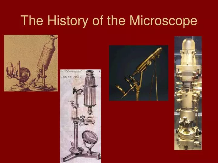
The History of the Microscope. Circa 1000AD – The first vision aid was invented (inventor unknown- possibly a monk) called a reading stone . It was a glass sphere that magnified when laid on top of reading materials.

E N D
Circa 1000AD – The first vision aid was invented • (inventor unknown- possibly a monk) called a reading stone. It was a glass sphere that magnified when laid on top of reading materials.
Circa 1284 - Italian, Salvino D'Armate is credited with inventing the first wearable eyeglasses
1590 – Two Dutch eye glass makers, Zaccharias Janssen and son Hans Janssen • experimented with multiple lenses placed in a tube. The Janssens observed that viewed objects in front of the tube appeared greatly enlarged, creating both the forerunner of the compound microscope and the telescope.
1665 – English physicist, Robert Hooke looked at a sliver of cork through a microscope lens and noticed some "pores" or "cells" in it.
1674 – Anton van Leeuwenhoek built a simple microscope with only one lens to examine blood, yeast, insects and many other tiny objects.
1830 – Joseph Jackson Lister reduces spherical aberration or the "chromatic effect" by showing that several weak lenses used together at certain distances gave good magnification without blurring the image. This was the prototype for the compound microscope.
1903 – Richard Zsigmondy developed the ultramicroscope that could study objects below the wavelength of light. He won the Nobel Prize in Chemistry in 1925.
1932 – Frits Zernike invented the phase-contrast microscope that allowed for the study of colorless and transparent biological materials for which he won the Nobel Prize in Physics in 1953.
1931 – Ernst Ruska co-invented the electron microscope for which he won the Nobel Prize in Physics in 1986. An electron microscope depends on electrons rather than light to view an object, electrons are speeded up in a vacuum until their wavelength is extremely short, only one hundred-thousandth that of white light. Electron microscopes make it possible to view objects as small as the diameter of an atom.
1981 – Gerd Binnig and Heinrich Rohrerinvented the scanning tunneling microscope that gives three-dimensional images of objects down to the atomic level. Binnig and Rohrer won the Nobel Prize in Physics in 1986. The powerful scanning tunneling microscope is the strongest microscope to date. STM image, 7 nm x 7 nm, of a single zig-zag chain of Cs atoms (red) on the GaAs(110) surface (blue). STM image, 35 nm x 35 nm, of single substitutional Cr impurities (small bumps) in the Fe(001) surface.
Power of the Electron Microscope If pushed to the limit, electron microscopes can make it possible to view objects as small as the diameter of an atom. Most electron microscopes used to study biological material can "see" down to about 10 angstroms--an incredible feat, for although this does not make atoms visible, it does allow researchers to distinguish individual molecules of biological importance. In effect, it can magnify objects up to 1 million times. Nevertheless, all electron microscopes suffer from a serious drawback. Since no living specimen can survive under their high vacuum, they cannot show the ever-changing movements that characterize a living cell.
Z-contrast scanning transmission electron microscope image (left) of iodine atoms in a carbon nanotube (visualized above).
Image of Si Atoms of n-type MoS2, a common dry lubricant. The bright spots indicate S atoms, which account for its excellent lubrication properties.
The letters “IBM” spelled in xenon atoms, as imaged by the atomic force microscope. Courtesy: IBM. In 1989, IBM scientist Don Eigler was surprised to learn that in addition to using an STM to look at tiny things he could also use it like a pair of tweezers, to move things as small as a single atom. That year, he used an STM to move individual atoms of the gas xenon that had been cooled to extremely low temperatures so that they sat still, to spell out “IBM” on a nickel surface. He then used the microscope to capture an image of the microscopic logo, and announced his results to the world.
Here’s an example of a question you might be asked on this material: • Which of the following is an instrument that a scientist might use to “see” an image of a molecule? A. a telescope • B. binoculars • C. electron microscope • D. anemometer
Sorry, a telescope is used to look at objects that are very far away such as stars and other planets • Try again
Sorry, binoculars are used to make things that you can see with your eyes appear closer • Try again
Sorry, an anemometer is used to measure wind speed • Try again
Congratulations!!!You got it!! • Yes!! An electron microscope can give us images that show us what molecules and even some atoms look like.