Download
1 / 23
280 likes | 356 Views
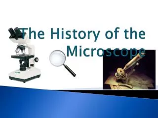
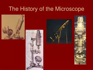
A Short History of the Microscope. Biology 20 Mrs. S. Pipke-Painchaud. http://www.cas.muohio.edu/~mbi-ws/microscopes/index.html. http://www.semguy.com/gallery.html. History ~ Inventors. Founding Fathers of Microscopy: http://www.cas.muohio.edu/~mbi-ws/microscopes/fathers.html

E N D
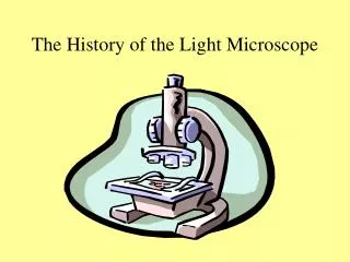
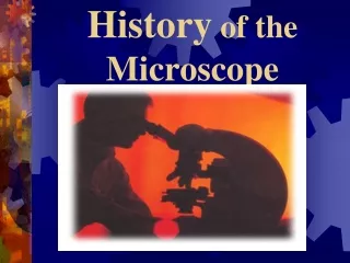
A Short History of the Microscope Biology 20 Mrs. S. Pipke-Painchaud
http://www.cas.muohio.edu/~mbi-ws/microscopes/index.html http://www.semguy.com/gallery.html
History ~ Inventors • Founding Fathers of Microscopy: http://www.cas.muohio.edu/~mbi-ws/microscopes/fathers.html • Please refer to the above website for information on the inventors of the microscope. (If the site is not available, please refer to the end of this powerpoint). • **Link to end of Powerpoint
Background • “The microscope was first built in 1595 by Hans and Zacharias Jansen (1588-1631) in Holland (see figure). • Later, it was perfected in the 17th century in several countries, including by Robert Hooke (1635-1703), in England but most notably by a Dutchman, Anton van Leeuwenhoek (1632-1723). • Hooke, after examining thin pieces of cork, discovered it had a honeycombed structure, and used for the first time the word "cell" to describe its smaller elements. Using a much improved microscope, with a monocular eyepiece, a wooden tube, a stage for holding a specimen, and a glass globe full of water to concentrate light onto it, Hooke produced marvelous illustrations, which were published in 1667, in his famous book Micrographia, which fired the imagination of his contemporaries, including van Leeuwenhoek.” • (Brain and Mind Website, Renato M.E. Sabbatini, PhD http://www.cerebromente.org.br/n17/history/neurons1_i.htm)
Hans and Zacharias Janssen, ~1590, Dutch Eyeglass Makers, Inventors • “Credit for the first microscope is usually given to Zacharias Janssen, pictured at the left, in Middleburg, Holland, around the year 1595. Since Zacharias was very young at that time, it's possible that his father Hans made the first one, but young Zach took over the production. • The first compound microscopes produced by the Janssen's was simply a tube with lenses at each end. The magnification of these early scopes ranged from 3X to 9X, depending on the size of the diaphragm openings.” Info directly from: Founding Fathers of Microscopy, http://www.cas.muohio.edu/~mbi-ws/microscopes/fathers.html
Background • 14th century lenses were used in spectacles • Late 16th century the Dutch refined the art of lens grinding significant magnification. • 1600s – lenses first mounted on permanent frameworks (so distance could be changed) • Why would this be important? • To focus the image
Next, lenses were paired together. • These formed the earliest compound microscopes and telescopes. • Why would this be useful? • To increase the magnification http://micro.magnet.fsu.edu/primer/museum/earlyitaliancompound1700s.html
Robert Hooke, 1635-1703,English Chemist, Mathematician, Physicist, and Inventor • “Hooke's remarkable engineering abilities enabled him to invent and improve many mechanical devices, including timepieces (for which he invented the spiral spring), the quadrant, and the Gregorian telescope. Perhaps even more intriguing than his actual inventions are the devices he designed but never built: he anticipated the invention of the steam engine, and as early as 1684 he described a working telegraph system. Hooke balanced his inventions with more pure research. Hooke improved on early compound microscopes around 1660.” Info directly from: Founding Fathers of Microscopy, http://www.cas.muohio.edu/~mbi-ws/microscopes/fathers.html
“In Micrographia (1665), he coined the word cell to describe the features of plant tissue (cork from the bark of an oak tree) he was able to discover under the microscope. He put his extensive mathematical knowledge in formulating the theory of planetary movement, which provided a basis for Sir Isaac Newton's theories of gravitation. In 1667 he discovered the role of oxygenation in the respiratory system.” Info directly from: Founding Fathers of Microscopy, http://www.cas.muohio.edu/~mbi-ws/microscopes/fathers.html
Illustration of Cork Cells by Robert Hooke http://askabiologist.asu.edu/research/buildingblocks/rhooke.html
Robert Hooke: http://www.fairfield.k12.ct.us/tomlinson/ctomlinson03/CellProject04/Per3/3OB/Q2.htm Robert Hooke's microscope. Hooke first described cells in 1665. http://www.biologyreference.com/Gr-Hi/History-of-Biology-Cell-Theory-and-Cell-Structure.html
Anton van Leeuwenhoek • refined lens grinding so that living things could be seen through the microscope. • Then there was little change until the industrial revolution Leeuwenhoek’s primitive one lens microscope. Both image from: http://www.cerebromente.org.br/n17/history/neurons1_i.htm
Anton van Leeuwenhoek, 1632-1723, Wine Assayer, Surveyor, Cloth Merchant, Minor Public Official, and Inventor • “Leeuwenhoek was a man with many talents, his most important attributes were creativity, power of observation, and ingenuity. Leeuwenhoek was a common man without any fortune or formal education, so he had to work for a living. Leeuwenhoek made simple (one lens) microscopes. He was not the first person to build a microscope, but the microscopes that he did build were the best ones for that time period. Leeuwenhoek was the first person to describe bacteria (from teeth scrapings), protozoans (from pond water), helped to prove the theory of blood circulation. He gained much of his inspiration form reading Hooke's Micrographia.” Info directly from: Founding Fathers of Microscopy, http://www.cas.muohio.edu/~mbi-ws/microscopes/fathers.html
“Van Leeuwenhoek used his new instrument, which was tenfold more potent than Hooke´s (he reached the amazing power of 300 times with a single lens!) to discover startling microscopic things, such as protozoa and spermatozoa, which thus far were completely unknown to science, or to discover the microscopic structure of known things, such as fleas and plant leaves. • Van Leeuwenhoek had even been able to slice specimens of a cow's optical nerves in 1674, and observe its longitudinal fibrous internal structure. He was perplexed to see that they were not hollow tubes, as the prevailing theory of the time, such as that defended by René Descartes, proposed.” • ((Brain and Mind Website, Renato M.E. Sabbatini, PhD http://www.cerebromente.org.br/n17/history/neurons1_i.htm) • View Bacterial drawings: • http://www.virtuallaboratory.net/Biofundamentals/labs/EColi%20introduced/section_01.html
Changes of the Industrial Revolution • standardized parts (which were interchangeable with other microscopes) lead to mass production • This triggered a drop in price increased access new discoveries clearer images • In approx. 1880 modern microscopes were being used
Electron Microscope • Developed in the 1930s • the electron microscope allowed for higher magnification • used electron beams (instead of light) and focused with an electromagnet (no lenses) • the light microscope produces magnifications up to 2000X • the electron microscope produces images that are magnified up to 50 000X or higher • What do you think the electron microscope allowed scientists to see? • Better quality images at higher magnification
Monocular Compound Microscope http://www.ascoindia.com/pcat-gifs/products-small/ms-351.jpg Binocular Compound Microscope http://www.labessentials.com/Rev3.jpg
Electron Microscope Termite Head: http://alfa.ist.utl.pt/~cvrm/staff/vramos/SIP.html http://www.phy.cuhk.edu.hk/centrallaboratory/CM120/CM120.html
Refer to this physics site for sample electron photos: • http://www.deutsches-museum.de/ausstell/dauer/physik/e_phys23.htm • View the SEM gallery of electron microscope images: • http://www.semguy.com/gallery.html
Relative Sizes • Refer to University of Arizona Website: • http://www.biology.arizona.edu/cell_bio/tutorials/cells/cells2.html
Parts of a Compound Microscope • Compound Microscope Parts: website reviews the parts of a microscope. (http://www.cas.muohio.edu/~mbi-ws/microscopes/microscopeparts.html) • Try the self test diagram http://biology.unm.edu/ccouncil/Biology_203/Summaries/ Microscopes.htm
Resource Sites: • University of Arizona: • Studying Cells: http://www.biology.arizona.edu/cell_bio/tutorials/cells/cells.html • Cell Video (animal and plant cells, image examples) • http://www.howe.k12.ok.us/~jimaskew/bvid4a.mov • http://www.howe.k12.ok.us/~jimaskew/unit4.htm