Download
1 / 44
500 likes | 724 Views
Ultrasound and Doppler Basics. Dr. Aarathy Appukuttan Final year resident Radiodiagnosis. Ultrasound imaging, also called sonography, involves exposing part of the body to high- frequency sound waves to produce images of the inside of the body.

E N D
Ultrasound and Doppler Basics Dr. Aarathy Appukuttan Final year resident Radiodiagnosis
Ultrasound imaging, also called sonography, involves exposing part of the body to high- frequency sound waves to produce images of the inside of the body. • Ultrasound examinations do not use ionizing radiation (as used in x-rays).
Because ultrasound images are captured in real- time, they can show the structure and movement of the bodys internal organs, as well as blood flowing through blood vessels.
Ultrasound is the most widely used imaging technology worldwide • Popular due to availability, speed, low cost, patient-friendliness (no radiation) • Applied in obstetrics, cardiology, inner medicine, urology
Can help to diagnose a variety of conditions and to assess organ damage following illness. • Is used to help physicians evaluate symptoms such as: • pain • swelling • infection • hematuria
heart and blood vessels • Liver • Gallbladder • Spleen • Pancreas • Kidneys • Bladder • Uterus and ovaries, • unborn child (fetus) in pregnant patients • Eyes • Thyroid and parathyroid glands• Scrotum • brain in infants• hips in infants
guide procedures such as needle biopsies, in which needles are used to extract sample cells from an abnormal area for laboratory testing. • image the breasts and to guide biopsy of breast cancer • diagnose a variety of heart conditions and to assess damage after a heart attack or diagnose for valvular heart disease.
Doppler ultrasound images can help the physician to see and evaluate: • blockages to blood flow (such as clots). • narrowing of vessels (which may be caused by plaque). • tumors and congenital vascular malformation
With knowledge about the speed and volume of blood flow gained from a Doppler ultrasound image, the physician can often determine whether a patient is a good candidate for a procedure like angioplasty.
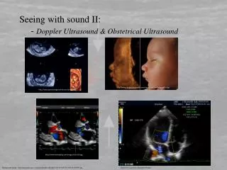
Applications in Obstetrics • Follow fetal development • Detect pathologies • Two-dimensional B-mode Ultrasound image and 3D image of a fetus
Applications in Cardiology • Blood flow in vessels • Contraction, Rhythm • Blood flow in the heart (defects on wall muscle, valve defects • Assessment of cardiac perfusion Prenatal diagnostic of the Fallot-Tetralogy
Applications in Musculoskeletal System • Visualisation of tendons, ligaments • Investigations under movement is possible – simplifies the detection of ruptures, obstructions
Applications of Ultrasound Elastography • US Elastography is often used to classify tumours. • Malignant tumours are 10 to 100 times stiffer than the normal soft tissue around Elastogram (of a breast) indication a mass with a high probability of being malignant tumour
Equipment • Ultrasound scanners consist of a console containing a computer and electronics, a video display screen and a transducer that is used to do the scanning. • The transducer is a small hand-held device that resembles a microphone, attached to the scanner by a cord.
The transducer sends out inaudible high frequency sound waves into the body and then listens for the returning echoes from the tissues in the body. • The principles are similar to sonar used by boats and submarines.
Properties of Ultrasound • The frequencies of medical Ultrasound waves are several magnitudes higher than theupper limit of → human hearing.
The ultrasound image is immediately visible on a video display screen that looks like a computer or television monitor. • The image is created based on the amplitude (strength), frequency and time it takes for the sound signal to return from the area of the patient being examined to the transducer and the type of body structure the sound travels through.
How is the procedure performed? • For most ultrasound exams, the patient is positioned lying face-up on an examination table that can be tilted or moved.
A clear water-based gel is applied to the area of the body being studied • To help the transducer make secure contact with the body and eliminate air pockets between the transducer and • The skin that can block the sound waves from passing into your body.
The sonographer (ultrasound technologist) or radiologist then presses the transducer firmly against the skin in various locations, sweeping over the area of interest or angling the sound beam from a farther location to better see an area of concern.
Doppler • Doppler ultrasound, a special application of ultrasound, measures the direction and speed of blood cells as they move through vessels. • The movement of blood cells causes a change in pitch of the reflected sound waves (called the Doppler effect). • A computer collects and processes the sounds and creates graphs or color pictures that represent the flow of blood through the blood vessels.
the Doppler effect is used to measure blood flow velocity. • Ultrasound reflected from red blood cells will change in frequency according to the blood flow velocity. • When direction of blood flow is towards the transducer, the echoes from blood reflected back to the transducer will have a higher frequency than the one emitted from the transducer.
When the direction is away from the transducer, the echoes will have a lower frequency than those emitted. • The difference in frequency between transmitted and received echoes is called the Doppler frequency shift, and this shift in frequency is proportional to the blood flow velocity.
DOPPLER FREQUENCY SHIFT • The Doppler shift is the difference between the incident frequency and reflected frequency. • When the reflector is moving directly away from or toward the source of sound, the Doppler frequency shift (fd) is calculated as where fI is the frequency of the sound incident on the reflector and fr is the frequency of the reflected sound. • Thus, the Doppler shift is proportional to the velocity of the blood cells.
TYPES OF DOPPLER OPERATION • 1. Continuous wave Doppler • 2. Pulsed wave doppler • 3. Duplex scanning • 4. Color flow doppler imaging • 5. Power dopple
What are the benefits ? • Most ultrasound scanning is noninvasive (no needles or injections) and is usually painless. • widely available, easy-to-use and less expensive than other imaging methods. • does not use any ionizing radiation.
Gives a clear picture of soft tissues that do not show up well on x-ray images. • The preferred imaging modality for the diagnosis and monitoring of pregnant women and their unborn babies. • Provides real-time imaging, making it a good tool for guiding minimally invasive procedures such as needle biopsies and needle aspiration
Limitations • Ultrasound waves are disrupted by air or gas; • Therefore ultrasound is not an ideal imaging technique for air-filled bowel or organs obscured by the bowel. • In most cases, barium exams, CT scanning, and MRI are the methods of choice in this setting.
Large patients are more difficult to image by usg because greater amounts of tissue attenuates (weakens) the sound waves as they pass deeper into the body. • Usg has difficulty penetrating bone and, therefore, can only see the outer surface of bony structures and not what lies within (except in infants). • For visualizing internal structure of bones or certain joints, other imaging modalities such as MRI are typically used.