Download
1 / 39
570 likes | 1.06k Views
Introduction to Carotid Ultrasound and Transcranial Doppler Ultrasound. Ryan Hakimi, DO, MS Director, Critical Care Neurology Assistant Professor Department of Neurology The University of Oklahoma Health Sciences Center January 16, 2015. Disclosures.

E N D
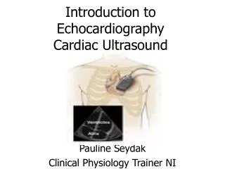
Introduction to Carotid Ultrasound and Transcranial Doppler Ultrasound Ryan Hakimi, DO, MS Director, Critical Care Neurology Assistant Professor Department of Neurology The University of Oklahoma Health Sciences Center January 16, 2015
Disclosures • Many of the slides have been adapted from slides presented at the American Society for Neuroimaging Annual Meetings by my mentors • FINANCIAL DISCLOSURE • Nothing to disclose • UNLABELED/UNAPPROVED USES DISCLOSURE • Nothing to disclose Andre Alexandrov, MD Zsolt Garami, MD Charles Tegeler, MD Alex Razumovsky, PhD
Objectives Review the basic principles of carotid ultrasound (CUS) and transcranial Doppler ultrasound (TCD) Illustrate the process of plaque morphology and assignment of range of carotid stenosis Illustrate the process of determining vasospasm by TCD Discuss some of the current applications of TCD
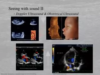
Principles of Ultrasound Blood flow velocity through a cross sectional area of a particular vessel (cm/s) Blood flow velocity is directly related to Doppler shift If you measure the Doppler shift you can derive the blood flow velocity Carotid ultrasound and transcranial Doppler ultrasound can accomplish this
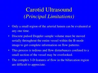
Carotid Ultrasound • Indications • Ischemic stroke or TIA • Assessment of carotid bruit • Assessment of carotid stenosis or occlusion • Pre-operative assessment for cardiovascular surgery • Post carotid endarterectomy or stenting
Principles of Carotid Duplex • Duplex (B-mode and Doppler) • B-mode (brightness mode) • Grayscale, used for visualization of structures and assessment of plaque morphology • Hyperechoic (bright white: bone, calcium), causes a shadow posterior to it • Hypoechoic (black or grey: thrombus) • Doppler velocities (peak systolic and end diastolic) • Used to determine the direction of blood flow • Used to estimate the range of stenosis • Each lab should have own validated parameters of the velocities to be used for assignment of stenosis, not just use published values
Assignment of Carotid Stenosis Carotid Artery Stenosis: Gray-Scale and Doppler US Diagnosis—Society of Radiologists in Ultrasound Consensus Conference Grant et al., 2003
Transverse Right ICA B-Mode Skin surface Fat and subcutaneous tissue internal jugular vein muscle common carotid artery (Transverse Right Proximal Common Carotid Artery)
Longitudinal Right ICA B-mode Plaque Focal irregular, heterogeneous plaque in the R Prx ICA (Longitudinal Right Proximal Internal Carotid Artery)
Right ICA B-mode with Color Doppler + Flow (toward probe) - Flow (away from probe) Patient’s Head Flow (away from probe) (Right Proximal Internal Carotid Artery)
Right ICA Doppler Velocities + Flow (away from probe) - Flow (away from probe) Flow (away from probe) Patient’s Head Cardiac irregularity (Right Proximal Internal Carotid Artery)
Report • Focal plaque in the right internal carotid artery • No hemodynamically significant stenosis demonstrated in the right internal carotid artery. • High resistance with focal plaque in the right internal carotid artery • Cardiac irregularity was noted
Longitudinal Left CCA B-mode Plaque Plaque with fibrous cap and lipid rich core Hyperechoic plaque with posterior acoustic shadowing (Longitudinal Left Mid Common Carotid Artery)
Longitudinal Left CCA B-mode Plaque Heterogeneous plaque with posterior acoustic shadowing (Longitudinal Left Distal Common Carotid Artery)
Longitudinal Left ICA Color Doppler Noise (sample volume picking up multiple velocity jets)
Carotid US vs Transcranial Doppler US • Carotid Ultrasound • Directly visualize the vessel • Stenosis determined by peak systolic velocity • TCD • Blind insonation • Uses mean velocity
Pulsatility Indices Low PI AVM low ejection fraction aortic regurgitation High PI: intracranial atherosclerosis increased intracranial pressure
Allows for evaluation of: Collateral flow Cerebral embolism Poor vasomotor reserve i.e. progression of carotid stenosis TCD use in Carotid Occlusive Disease
TCD use in Carotid Occlusive Disease Slide courtesy Z. Garami, MD
Risk of Ipsilateral Stroke: Number of Activated Collaterals (ACA, PCOM, OA)
Subarachnoid Hemorrhage Detection of vasospasm Clinical exam (usually somnolence or non-focal symptoms), not very sensitive Daily TCD (non-invasive, 90% sensitivity, often precedes clinical vasospasm) TCD or CTA can be used to screen for “plasty-able” lesions Images from 1. http://www.spencertechnologies.com/products.html#thumb 2. www.viswiki.com/en/Transcranial_doppler, 3. http://depts.washington.edu/uwtcdlab/images/tcd/tcd3_lg.gif all accessed on 1/24/2010 4. Cerebrovascular Ultrasound in Stroke Prevention and Treatment
Thresholds for Anterior Circulation Vasospasm Should be validated at the given center Mild vasospasm 120-140 cm/s, Lindegaard Ratio (MCA velocity/extracranial ICA) 3-4 Moderate vasospasm 140-180 cm/s, Lindegaard Ratio (MCA velocity/extracranial ICA) 4-6 Severe vasospasm >200 cm/s, Lindegaard Ratio (MCA velocity/extracranial ICA) >6 Less reliable in posterior circulation (due to greater anatomic variance)
Data on TCD Monitoring Courtesy Mauro Oddo, MD
Subarachnoid Hemorrhage Triple-H therapy (hypertension, hypervolemia, hemodilution) Introduced in 1970’s to prevent delayed cerebral ischemia from vasospasm Prophylactic triple-H (in absence of vasospasm) does not prevent vasospasm (Treggiari et al. J. Neurosurg 2008) Double-H therapy (hypertension and hypervolemia as hemodilution is consequence of hypervolemia) Must be individualized and titrated to clinical exam and TCD Can result in pulmonary edema, hyponatremia, MI, etc.
Brain Death • Uniform Determination of Death Act • Legally acknowledged brain death as a mechanism of death • Defined death as: • Irreversible cessation of circulatory and respiratory functions OR • Irreversible cessation of all functions of the entire brain, including the brain stem 1. Guidelines for the determination of death: report of the medical consultants on the diagnosis of death to the President’s commission on the ethical problems in medicine and biochemical and behavioral research. JAMA 1981;246:2184-2186. 2. Uniform Determination of Death Act, 12 uniform laws annotated 589 (West 1993 West suppl 1997)
AAN recommendations on use of ancillary testing AAN Clinician Guideline Supplement: Ancillary Testing; Update: Determining Brain Death in Adults. 2010.
Diagnosis of Patent Foramen Ovale (PFO) Slide courtesy Alex Razumovsky, PhD
Diagnosis of Patent Foramen Ovale (PFO) • PFO is a residual channel between the right and left atrium which originally allowed oxygenated placental blood to pass from the right to left atrium bypassing the fetal lungs • Usually closes by age 2, but can persist in 25-30% of the general population
Diagnosis of PFO by TCD • Agitated saline study • Monitor the MCA Slide courtesy Z. Garami, MD
Diagnosing subclavian steal by TCD • Image the vertebral artery • Ischemic cuff test
Subclavian Steal Syndrome Slide courtesy Z. Garami, MD
OU Neurology Questions Thank you