Download
1 / 22
220 likes | 438 Views
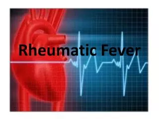
Rheumatic Fever. Normal Heart Anatomy. Rheumatic Fever (RF). Definition: Rheumatic fever (RF) is an autoimmune disease affecting the heart and extra- cardiac sites (joints, brain, skin and others).

E N D
Rheumatic Fever (RF) Definition: • Rheumatic fever (RF) is an autoimmune disease affecting the heart and extra- cardiac sites (joints, brain, skin and others)
The incidence of RF has been lowered in the developed countries but is still high in poor communities • The disease affects children and young adults (5-15years) • The disease follows upper respiratory infection (tonsillitis) with Group A Beta hemolytic streptococci
Theories of Pathogenesis: • Toxic products of streptococci • Immunologic cross-reactivity between Streptococcal substances and heart muscle (heart reactive antibodies) • Sensitized T-lymphocytes may lead to cardiac injury
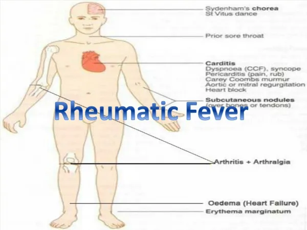
Major Manifestations Carditis (friction rub, murmur, cardiomegaly, CHF) Arthritis (migratory polyarthritis, swollen, red, tender) Chorea Subcutaneous nodules Erythema marginatum Minor Manifestations Clinical Fever Arthralgia History of rheumatic fever or rheumatic heart disease Laboratory Acute phase reactants (ESR, C-reactive protein, leukocytosis) Prolonged P-R interval on ECG JONES' CRITERIA FOR DIAGNOSIS OF RF:
PATHOLOGY OF RHEUMATIC FEVER • Cardiac Disease (Rheumatic heart disease) • Extra-Cardiac Disease
RHEUMATIC HEART DISEASE • Rheumatic heart disease: all the heart layers are affected (pancarditis) • Rheumatic myocarditis • Rheumatic pericarditis • Rheumatic endocarditis
1- Rheumatic myocarditis: Acute phase: it is characterized by the development of pathognomonic lesions called Aschoff’s Bodies within the myocardium. Gross features: • Aschoff bodies are multiple tiny nodules (1-2 mm in diameter) Microscopic features: • Aschoff body is a lesion composed of: • Fibrinoid necrosis ( destroyed fragmented collagen) • Surrounded by lymphocytes and histiocytes & • Aschoff cells (large mononuclear or multinuclear macrophages)
Aschoff’s body Blood vessel fibrinoid degeneration Aschoff cells
Chronic phase: • Over years or decades the Aschoff bodies undergo fibrous scarring
2- Rheumatic Pericarditis: • Acute phase: Aschoff bodies are formed accompanied by serofibrinous inflammation. • Chronic phase: Fibrosis and adhesions may occur between the visceral and the parietal layers of the pericardium
3- Rheumatic Endocarditis: It affects both mural and valvular endocardium • Mural Endocardium: • i- Acute phase: Aschoff bodies develop in the endocardium • ii- Chronic phase: healing results in a white patch
Valvular Endocardium • Vegetations (thrombi) develop at the lines of contact of the cusps causing friction of the swollen cusps.
Rheumatic Mitral Valve Small vegetations are formed at injured parts
CHRONIC RHEUMATIC VALVULAR DISEASE • Mitral & Aortic Valves Pathology: • Thickening of valve leaflet, especially along the lines of closure • Fusion of commissures • Result is mitral or aortic stenosis, insufficiency, or both
Rheumatic Mitral Stenosis Fusion of commisures Thick valve leaflet
EXTRACARDIAC LESIONS OF RHEUMATIC FEVER • Joints: Rheumatic arthritis affect the large joints in a fleeting way i.e joint inflammation is followed by joint resolution, then another joint become inflamed followed by resolution and so on. The affected joint is painful, tender, hot & swollen. • Brain: Rheumatic chorea (rapid involuntary purposeless movements); it is due to inflammation of the basal ganglia. The condition is reversible • Skin: Rheumatic subcutaneous nodules occur over bony prominences and their structure is similar to the Aschoff bodies. • Rheumatic arteritis: affecting the coronaries, renal, mesenteric and cerebral arteries • Pleurisy and peritonitis: serofibrinous type
PERICARDIAL DISEASES I. PERICARDITIS Inflammation of the pericardium • Causes • MI, Staphylococcus, tumor, TB, uremia
II. PERICARDIAL EFFUSION • Serous fluid in pericardial sac • Usual cause: Chronic Heart Failure
III. HEMOPERICARDIUM • Myocardial rupture from MI • Trauma • Bleeding from infection, tumor, etc. • Haemorrhage from aorta