Download

1 / 22
240 likes | 373 Views
Rheumatic Fever and Rheumatic Heart Disease. Dr. Tarek Atia. Acute Rheumatic Fever. Autoimmune consequence of pharyngeal infection with Group-A Beta Hemolytic Streptococci (GABHS). Generalized inflammatory response affecting joints , skin , subcutaneous tissues & the heart.

E N D
Rheumatic Fever and Rheumatic Heart Disease Dr. Tarek Atia
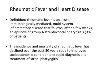
Acute Rheumatic Fever Autoimmune consequence of pharyngeal infection with Group-A Beta Hemolytic Streptococci (GABHS). Generalized inflammatory response affecting joints, skin, subcutaneous tissues & the heart. Modified Duckett-Jones criteria form the basis of the diagnosis of the condition.
About 66% of the patients with an acute rheumatic fever have a history of an upper respiratory tract infection several weeks before. • Acute rheumatic fever typically develops 2-4 weeks after an acute attack of GABHS pharyngitis at a time when clinical findings of pharyngitis are no longer present. • The peak age (6-15 yrs.) & seasonal incidence of acute rheumatic fever closely parallel those of GABHS infections. Acute Rheumatic Fever
Features suggestive of GABHS infection Patient 6 to 15 years of age Presentation in winter or early spring Fever, Headache Sudden onset of sore throat Nausea, vomiting & abdominal pain and pain with swallowing Swollen, red uvula Soft palate petechial hemorrhage. Tender, enlarged anterior cervical lymph nodes Tonsillo-pharyngeal erythema & exudates
Redness & swelling of throat & tonsils; Swollen, red uvula; Soft palate petechiae (“doughnut lesions”) Tonsillopharyngeal erythema & exudates Acute Rheumatic Fever Sore throat: fever, white draining patches on the throat & swollen or tender lymph glands in the neck
Supporting evidences: Patients with acute rheumatic fever almost always have serologic evidence of a recent GABHS infection Their antibody titers are usually considerably higher than those in patients with GABHS infections without acute rheumatic fever Antimicrobial therapy against GABHS prevents initial symptoms of acute rheumatic fever Long-term, continuous prophylaxis: prevents recurrences of acute rheumatic fever.
Predisposing factors Family history of rheumatic fever Low socio-economic status (poverty, poor hygiene, medical care deficiency) Age: 6-15 years
Pathogenesis: • GABHS pyogenes has a cell wall contain M-protein that are highly antigenic. • The antibodies generated against the M-protein may cross react with cardiac myofiber protein myosin, heart muscle glycogen and smooth muscle cells of arteries, releasing cytokine and tissue destruction. • Characteristic Aschoff bodies, composed of swollen eosinophilic collagen surrounded by lymphocytes and macrophages can be seen on light microscopy. The larger macrophagesmay become Aschoff giant cells.
Clinical Manifestations • Duckett-Jones in 1944 proposed guidelines to aid in diagnosis • There are 5 major and 4 minorcriteria with requirement for evidence (microbiologic or serologic) of recent GABHS infection. • According to Jones criteria, the diagnosis of rheumatic fever can be made when 2 major criteria or 1 major & 2 minor criteria along with the absolute requirement.
R R P P Prolonged PR interval
MAJOR MANIFESTATIONS 1- Migratory Polyarthritis • Most common (75%) • Involves larger joints: the knees, ankles, wrists & elbows • Rheumatic joints are hot, red, swollen & exquisitely tender • The joint involvement is characteristically migratory in nature • Monoarticular arthritis is unusual unless anti inflammatory therapy is initiated prematurely, aborting the progression of the migratory polyarthritis
2- Carditis • Occurs in 50% of patients • The most serious manifestations of acute rheumatic fever • Rheumatic carditis: pan-carditis with active inflammation of myocardium, pericardium & endocardium • Acute rheumatic carditis:tachycardia out of proportion to fever & cardiac murmurs. • Consists of either isolated mitral valvular disease or combined with aortic valvular disease
The major consequence of acute rheumatic carditis is chronic, progressive valvular disease:- • Mitral and /or aortic valveregurgitation orstenosis • However, in developing countries, where acute rheumatic fever often occurs at a earlier age, mitral stenosis & aortic stenosis may develop in young children • Moderate to severe rheumatic carditis is associated with cardiomegaly and congestive heart failure with hepatomegaly & peripheral & pulmonary edema • Other clinical finding; pericardial effusion, decreased ventricular contractility
3- Chorea • Affects 10-15% of patients, mainly girls (8-12 yrs) • Neuropsychiatric disorder: choreic movement, hypotonia & hyperactivity • Psychiatric signs: Begins with personality changes (poor school performance) • Replace in 1-4 weeks by characteristic spontaneous, purposeless movement of chorea (lasts 4-8 months) followed by motor weakness • Exacerbation by stress & disappearing with sleep are characteristic • Elevated titers of “antineuronal antibodies” have been found in over 90% of patients
4- Erythema Marginatum • A rare, affects < 3% of patients with acute rheumatic fever, but characteristic rash of acute rheumatic fever. • It consists of erythematous,macular lesions with pale centers that are not pruritic • It occurs primarily on the trunk & extremities, not on the face & it can be accentuated by warming the skin
5- Subcutaneous Nodules • A rare, affects ≤1% of patients with acute rheumatic fever. • Consist of firm nodules approximately 1cm in diameter along the extensor surfaces of tendons near bony prominences • A correlation between the presence of these nodules & significant rheumatic heart disease
MINOR MANIFESTATIONS Clinical: • Arthralgia (in the absence of polyarthritis as a major criterion) • Fever(typically temperature ≥39.5C & occurring early in the course of illness) Laboratory minor manifestations: • Elevated acute-phase reactants:C-reactive protein, erythrocyte sedimentation rate (ESR), polymorphonuclearleucocytosis. • Prolonged PR interval on electrocardiogram (1st degree heart block)
ESSENTIAL CRITERIA An absolute requirement for the diagnosis of acute rheumatic fever is supporting evidence of a recent GABHS infection
TREATMENT • Bed rest • Antibiotic Therapy: • 10 days of orally administered penicillin or erythromycin or a single intramuscular injection of benzathine penicillin to eradicate GABHS from the upper respiratory tract • Afterwards, the patient should be started on long-term antibiotic prophylaxis