Download
1 / 44
470 likes | 684 Views
Rheumatic Fever AND RHD. Dr.Abdulelah Mobeirek ( FRCPc ). Rhuematic Fever. Follows group A beta hemolytic streptococcal throat infection It represents a delayed immune response to infection with manifestations appearing after a period of 2-4 weeks Age 5-15 yrs A multisystem disease

E N D
Rheumatic Fever AND RHD Dr.AbdulelahMobeirek(FRCPc)
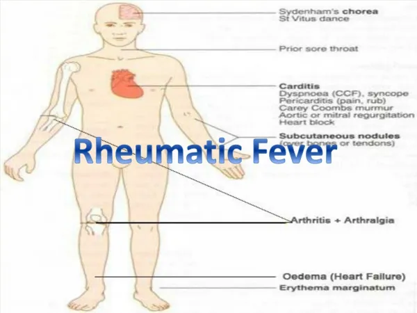
Rhuematic Fever • Follows group A beta hemolytic streptococcal throat infection • It represents a delayed immune response to infection with manifestations appearing after a period of 2-4 weeks • Age 5-15 yrs • A multisystem disease • Major effect on health is due to damage to heart valvves
Rheumatic Fever • A disease of poverty and low socioeconomic status • Rare in wealthy countries, due to improved living conditions, less overcrowding, and better hygiene with reduction in transmission of GABHS • Incidence: In Cambodia 2.2/1000 children, Mozambique 2.3/1000, India 0.75/1000
Global Burden of RHD • Total cases with RHD:20 Millions • CHF: 3 Million • Valve surgery required in 1 Million • Annual incidence of RF: 0.5 Million, nearly half develop carditis • Estimated deaths from RHD: 230,000/YR • Imposes a substantial burden on health care systems with limited budgets
Pathologic Lesions • Ashcoff nodules: • Fibrinoid degeneration of connective tissue, inflammatory cells
Clinical Features • Onset of acute rheumatic fever is typically characterized by an acute febrile illness 2 to 4 weeks after an episode of pharyngitis. • Diagnosis is primarily clinical and is based on a constellation of signs and symptoms, which were initially established as the Jones criteria
Modified Jones Criteria Major manifestations • Carditis • Polyarthritis • Chorea • Erythema marginatum • Subcutaneous nodules
Modified Jones Criteria Minor manifestations • Fever • Arthralgias • Previous H/O RF OR RHD • ↑CRP or ESR • Prolonged PR interval on ECG
Modified Jones Criteria Evidence of antecedent GABHS • Positive throat culture or rapid antigen test positive for GABHS • ↑ ASO Titer
Arthritis • Most common feature: present in 80% • Earliest manifestation of ARF • Major joints: The knees and ankles, shoulders, elbows • “Migrating”, “Fleeting” polyarthritis • Duration short < 1 week • Responds well to Salicylates • Does not progress to chronic disease
Sydenham Chorea • Occur in 5-10% of cases • Abrupt Purposeless involuantry movements of muscles of face, neck, trunk, and limbs. • May appear even 6 months after the attack of rheumatic fever • Clinically manifest as-clumsiness, deterioration of handwriting,emotional lability or grimacing of face
Subcutaneous Nodules • Occur in 10% • Usually 0.5 – 2 cm long • Firm non-tender • Occur over extensor surfaces of joints, on bony prominences, tendons, spine • Short lived: last for few days • Associated with severe carditis
Erythema Marginatum • Present in 5% • Reddish border, pale center, round or irregular serpiginous borders, non-pruritic, transient rash • Occurs on trunk, abdomen or proximal limbs • Associated with carditis
Erythematous patches with central clearing Erythema marginatum
Carditis • Occurs in40- 50% of cases • Pancarditis • Only manifestation of ARF that leaves permanent damage • Murmurs of MR or AR may occur in acute stage while mitral stenosis occurs in late stages • Cardiomegaly and CHF may occur
Modified Jones Criteria for Diagnosis of Acute Rheumatic Fever A firm diagnosis requires • 2 Major manifestations or 1 Major and 2 Minor manifestations and 2 ) Evidence of a recent streptococcal infection. However, when chorea or carditis is clearly present, evidence of an antecedent group A streptococcal infection is not necessary.
Differential Diagnosis • Juvenile rheumatiod arthritis • Infective endocarditis • Sickle cell arthropathy • Lupus • Myocarditis • Reactive arthritis • Leukemia
Investigations • CBC, ESR, CRP • Anti-streptococcal antibodies: ASO titre • Throat swab for culture • ECG • CXR • ECHO
Treatment of ARF • Salicylates : Aspirin • 75-100 mg /kg/day given as 4 divided doses for 6 -8 weeks • Attain a blood level 20-30 mg/dl • Prednisolone: • 2mg/kg/day taper over 6 weeks • Given when there is carditis
Treatment • Bed rest • Treat heart failure if present • Valve replacement later in life once symptoms develop or LV dysfunction occurs from severe valve regurgitation or valve stenosis
Secondary Prevention of Rheumatic Fever (Prevention of Recurrent Attacks) Agent Dose Mode Benzathine penicillin G 1 200 000 U every 4 weeks* Intramuscular or Penicillin V 250 mg twice daily Oral or Sulfadiazine 0.5 g once daily for patients 27 kg (60 lb Oral 1.0 g once daily for patients >27 kg (60 lb) For individuals allergic to penicillin and sulfadiazine Erythromycin 250 mg twice daily Oral *In high-risk situations, administration every 3 weeks is justified and recommended
Duration of Secondary Rheumatic Fever Prophylaxis CategoryDuration Rheumatic fever with carditis and 10 y since last episode residual heart disease or until age 40y ,(which- (persistent valvar disease*) ever is longer), sometimes life long prophylaxisRhumatic fever with carditis 10 yrs or until age 21yrs But no residual VHD (whichever is longer) Rheumatic fever without carditis 5 y or until age 21 y, ( whichever is longer) *
Mitral Stenosis • The normal MVA= 4-6 cm • In severe ms <1 • High LAP • The rise in LAP causes a similar rise in pulmonary capillaries, veins and artery
Mitral Stenosis • Long Asymptomatic period after initial attack of RF until onset of class 1 / 2 symptoms: 10 – 30 yrs ( latent period ) • Once symptoms develop there is another plateau of 5 –10 yrs before onset of AF • This followed by a period of 5-10 yrs until onset of class III- Iv symptoms
Clinical Features • Dyspnea • Fatigue • Palpitation • Hemoptysis (10%) • Hoarseness ( Ortner’s syndrome) • Dysphagia • Storoke or peripheral embolization
Clinical Features • Cyanosis (Mitral facies,malar flush) • Tapping apex ( S1) • Parasternal heave • Diastolic thrill • Accentuated S1 , accentuated S2 • Opening snap • Mid-diastolic rumble
Investigations • CXR Straightening of the left heart border Double density Kerley B lines , CA in MV • ECG: LAE, P Mitrale , RV dominance • ECHO: MVA, PAP
Management • B-Blockers ,CCB • Digoxin ( AF ) • Warfarin • Balloon Valvuloplasty • Mitral valve replacement
Mitral Regurgitation • Asymptomatic • Dyspnea , orthopnea, PND • Displaced PMI, Thrill • Soft S1, • Pansystolic murmur • Treatment is surgical
Aortic Regurgitation • Water-hammer / collapsing pulse • Wide pulse pressure • Corrigan’s sign • De Musset sign • Muller sign • Quincke’s pulse • Hill’s sign
Symptoms • Angina • Syncope • Dyspnea
Signs • Arterial Pulse wave form : plateau Small { Parvus } Slow rise { Tardus } • Sustained not displaced PMI • Systolic thrill • S4
Signs • Late peaking of murmur • Single S2 : Soft or absent A2 • Paradoxical splitting of S2
Aortic Valve Disease • Treatment is surgical: Aortic valve Replacement