Download
1 / 1
10 likes | 18 Views
Time Courses of fMRI Activations Spanning The Stimulus-Response Interval During Word Generation By Nonfluent Aphasics

E N D
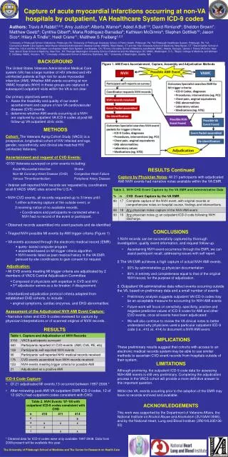
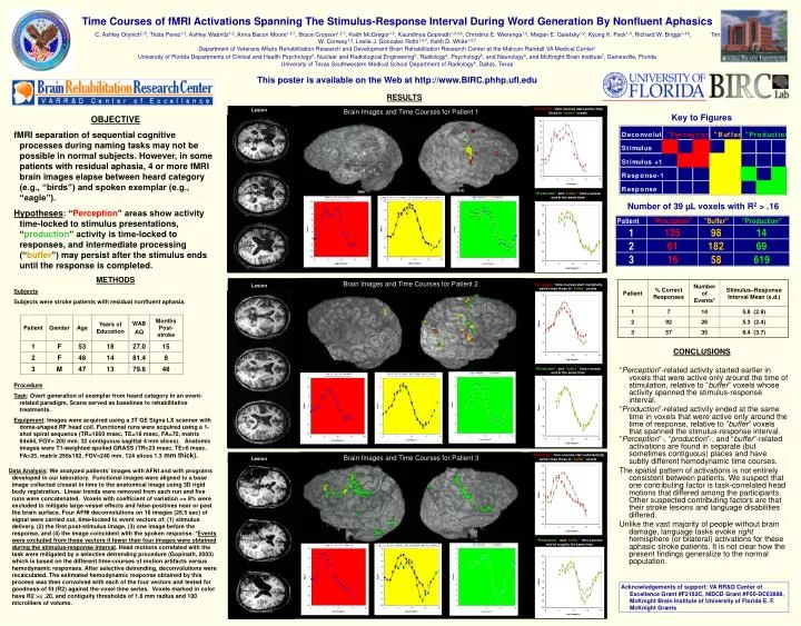
Time Courses of fMRI Activations Spanning The Stimulus-Response Interval During Word Generation By Nonfluent Aphasics C. Ashley Orynich1,5, Trista Perez1,5, Ashley Wabnitz1,2, Anna Bacon Moore1,2,7, Bruce Crosson1,2,7, Keith McGregor1,5, Kaundinya Gopinath1,3,4,8, Christina E. Wierenga1,2, Megan E. Gaiefsky1,2, Kyung K. Peck1,4, Richard W. Briggs1,4,8, Tim W. Conway1,2, Leslie J. Gonzalez Rothi1,6,7, Keith D. White1,5,7 Department of Veterans Affairs Rehabilitation Research and Development Brain Rehabilitation Research Center at the Malcom Randall VA Medical Center1 University of Florida Departments of Clinical and Health Psychology2, Nuclear and Radiological Engineering3, Radiology4, Psychology5, and Neurology6, and McKnight Brain Institute7, Gainesville, Florida University of Texas Southwestern Medical School Department of Radiology8, Dallas, Texas This poster is available on the Web at http://www.BIRC.phhp.ufl.edu RESULTS Lesion Brain Images and Time Courses for Patient 1 “Perception”time courses start earlier than those in“buffer”voxels Key to Figures • OBJECTIVE • fMRI separation of sequential cognitive processes during naming tasks may not be possible in normal subjects. However, in some patients with residual aphasia, 4 or more fMRI brain images elapse between heard category (e.g., “birds”) and spoken exemplar (e.g., “eagle”). • Hypotheses: “Perception” areas show activity time-locked to stimulus presentations, “production” activity is time-locked to responses, and intermediate processing (“buffer”)may persist after the stimulus ends until the response is completed. • METHODS • Subjects • Subjects were stroke patients with residual nonfluent aphasia. • Procedure • Task: Overt generation of exemplar from heard category in an event-related paradigm. Scans served as baselines to rehabilitative treatments. • Equipment: Images were acquired using a 3T GE Signa LX scanner with dome-shaped RF head coil. Functional runs were acquired using a 1-shot spiral sequence (TR=1660 msec, TE=18 msec, FA=70, matrix 64x64, FOV= 200 mm, 32 contiguous sagittal 4 mm slices). Anatomic images were T1-weighted spoiled GRASS (TR=23 msec, TE=6 msec, FA=25, matrix 256x192, FOV=240 mm, 124 slices 1.3 mm thick). “Production”and“buffer ” time courses end at the same time Number of 39 µL voxels with R2 > .16 Brain Images and Time Courses for Patient 2 “Perception”time courses start marginally earlier than those in“buffer”voxels Lesion CONCLUSIONS “Production”and“buffer ” time courses end at the same time “Perception”-related activity started earlier in voxels that were active only around the time of stimulation, relative to “buffer” voxels whose activity spanned the stimulus-response interval. “Production”-related activity ended at the same time in voxels that were active only around the time of response, relative to “buffer” voxels that spanned the stimulus-response interval. “Perception”-, “production”-, and “buffer”-related activations are found in separate (but sometimes contiguous) places and have subtly different hemodynamic time courses. The spatial pattern of activations is not entirely consistent between patients. We suspect that one contributing factor is task-correlated head motions that differed among the participants. Other suspected contributing factors are that their stroke lesions and language disabilities differed. Unlike the vast majority of people without brain damage, language tasks evoke right hemisphere (or bilateral) activations for these aphasic stroke patients. It is not clear how the present findings generalize to the normal population. Brain Images and Time Courses for Patient 3 “Perception”time courses start substantially earlier than those in“buffer”voxels Lesion Data Analysis: We analyzed patients’ images with AFNI and with programs developed in our laboratory. Functional images were aligned to a base image collected closest in time to the anatomical image using 3D rigid body registration. Linear trends were removed from each run and five runs were concatenated. Voxels with coefficient of variation >= 8% were excluded to mitigate large-vessel effects and false-positives near or past the brain surface. Four AFNI deconvolutions on 16 images (26.5 sec) of signal were carried out, time-locked to event vectors of: (1) stimulus delivery, (2) the first post-stimulus image, (3) one image before the response, and (4) the image coincident with the spoken response. *Events were excluded from these vectors if fewer than four images were obtained during the stimulus-response interval. Head motions correlated with the task were mitigated by a selective detrending procedure (Gopinath, 2003) which is based on the different time-courses of motion artifacts versus hemodynamic responses. After selective detrending, deconvolutions were recalculated. The estimated hemodynamic response obtained by this process was then convolved with each of the four vectors and tested for goodness-of fit (R2) against the voxel time series. Voxels marked in color have R2 >= .20, and contiguity thresholds of 1.8 mm radius and 100 microliters of volume. “Production”and“buffer ” time courses end at roughly the same time Lesion Acknowledgements of support: VA RR&D Center of Excellence Grant #F2182C, NIDCD Grant #P50-DC03888, McKnight Brain Institute of University of Florida E. F. McKnight Grants