Download
1 / 62
660 likes | 782 Views
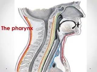
PHARYNX. Dr. Jamila -ELmedany. EXTENSION. It is a fibro muscular tube that lies behind: Nose . Mouth. Larynx. It extends from : Base of the skull To 6 th cervical vertebra. SHAPE. It is Funnel shaped. It has: Upper expanded end that lies under the skull and

E N D
PHARYNX Dr. Jamila -ELmedany
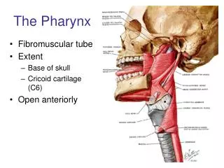
EXTENSION • It is a fibro muscular tube that lies behind: • Nose. • Mouth. • Larynx. • It extends from : Base of the skull • To • 6th cervical vertebra.
SHAPE • It is Funnel shaped. • It has: • Upper expanded end that lies under the skull • and • A narrow lower end • that becomes continuous with the Esophagus.
RELATIONS • Posterior: • Prevertebral muscles and fascia. • Lateral: • (1) Auditory tube. • (2) Styloid process and its muscles.
RELATIONS • (3) Carotid sheath and its contents. • (4) Thyroid gland.
ANTERIOR RELATIONS • It is deficient. • It is replaced by: • Posterior nasal openings. • Oropharyngeal isthmus. • Inlet of the larynx.
STRUCTURE OF THE WALL • 1. Mucosa : • Continuous with that of the nose ,auditory tube, oral cavity , larynx and esophagus. • 2. Fascia: Thickened • Internally: Paryngobasilar fascia. • Externally: Buccopharyngefascia. • 3. Muscles :circular & longitudinal.
CIRCULAR MUSCLES • CONSTRICTORS: • Superior. • Middle. • Inferior. • They propel the bolus of food down into the esophagus. • They overlap like three glasses stacked one inside the other.
CONSTRICTORS • Each one fans out from its anterior attachment and passes posteriorly around the pharynx. • They join each other in the: • Pharyngeal Raphe • A fibrous midline raphe which extends from the occipital bone to the esophagus.
CRICOPHARYNGEUS • It is the lowest fibers of the inferior constrictor muscle. • Action : • 1. A sphincter on the lower end of the pharynx. • 2. Prevents the entry of air into the esophagus in between swallowing.
LONGITUDINAL MUSCLES • Stylopharyngeus. • Salpingopharyngeus. • Palatopharyngeus. • Origins : • Styloid process . • Pharyngotympanic tube. • Soft palate.
LONGITUDINAL MUSCLES • Insertion: • To the pharyngeal wall. • Action: • 1. Elevate the larynx and pharynx. • 2. Pull the pharynx forward during swallowing and speaking.
STRUCTURES THROUGH GAPS • (1) Between: • Base of the skull and the superior constrictor : • Pharyngobasilar fascia. • Tensor palati. • Levator palati.
STRUCTURES THROUGH GAPS • (2) Between: • Superior and Middle constrictors: • It is a triangular gape . • It allows : • A. muscles, vessels & nerves to pass between the tongue and regions lateral to pharynx. • B. Stylopharyngeustendon to slip into the pharynx.
STRUCTURES THROUGH GAPS • (3) Between: • Middle & Inferior constrictors: • Internal laryngeal nerve & superior laryngeal vessels. • (4) Below: • Inferior constrictor: • Therecurrent laryngealnerve &inferior laryngeal vessels.
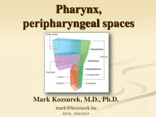
NASOPHARYNX • It has a respiratory function. • It is Behind the posterior nasal openings (Choanae) • AND • Above the Soft palate.
BOUNDARIES • Roof: • Basilar part of occipital bone. • Body of sphenoid. • It has • A collection of lymphoid tissue • (pharyngeal tonsil) in its submucosa.
BOUNDARIES • Floor : • Sloping upper surface of the soft palate. • Posterior wall: • Anterior arch of atlas. • Anterior : • Choanae.
PHARYNGEAL ISTHMUS • It is a gap in the floor between the free end of the soft palate and the posterior wall. • It is closed in swallowing by the elevation of the soft palate and pulling forward of the posterior wall.
BOUNDARIES • Lateral wall : • 1.Pharyngealopeningof the auditory tube. • 2.Tubal elevation: • The elevated posterior margin of the tube. • Pharyngeal Recess : • It is a slit like depression on each side behind the tubal elevation.
LATERAL WALL • 3. Salpingo-pharyngealfoldof mucous membrane produced by the salpingopharyngeal muscle. • 4.Tubal tonsil: • It is a collection of lymphoid tissue behind the opening of the auditory tube.
ADENOID • Hypertrophy and infection of the pharyngeal tonsils. • It causes: obstruction of the posterior nasal openings. • The patient: breathes through the mouth and snores loudly at night. • It can cause: deafness and recurrent otitis media due to its close relation to the auditory tube.
OROPHARYNX • It is behind the mouth cavity. • It extends from: • Soft palate • TO • The upper border of the Epiglottis.
BOUNDARIES • Roof : • It is the under surface of the soft palate and the pharyngeal isthmus. • It has collection of lymphoid tissue.
BOUNDA RIES • Floor : • Posterior 1/3 of the tongue & the interval between the tongue and the anterior surface of the epiglottis. • Lingual tonsil : • A collection of lymphoid tissue on the posterior 1/3 of the tongue.
BOUNDARIES • Lateral wall : • Palatoglossal arch • A mucous fold overlying the palatoglossal muscle. • It marks the boundary between the oral cavity and the oropharynx. • The arched opening between the two folds is the Oropharyngeal isthmus.
LATERAL WALL • Palato-pharyngeal arch • A mucous fold behind the palatoglossal arch. • It overlies the palatopharyngeal muscle.
BOUNDARIES • Anterior : • It opens into the mouth cavity through theoropharyngeal isthmus. • Posterior wall : • Body of 2nd cervical vertebra. • The upper part of the body of the 3rd vertebra.
PALATINE TONSILS • Two oval bodies of lymphoid tissue. • They have their maximum size in childhood. • They lie on each side in the tonsilar sinusin thelateral wall of the oropharynx between the palatoglossal arches (anterior) and palatppharyngeal arches(posterior)
PALATINE TONSILS • They lie on the mucosa lining the Superior Constrictor muscle. • The capsule is separated from the superior constrictor muscle by loose fascia.
PALATINE TONSILS • This fascia contains: • The External palatine vein. • The Facial artery is lateral to the muscle. • The Internal carotid arterylies about 1’’ (2.5) cm behind and lateral to the tonsil.
BOUNDARIES • Superior: • Soft palate. • It is continuous with the lymphoid tissue on its undersurface . • Inferior : • Posterior third of tongue. • It is continuous with the lingual tonsil.
BLOOD SUPPLY • Arterial : • Tonsillar arteries from : • Facial. • Ascending palatine. • Venous Drainage: External palatine vein. Pharyngeal venous plexus. • Pharyngeal and Facial veins.
LYMPH DRAINAGE • Juglodigastricnodes: • Below and behind mandible.
SENSORY SUPPLY • Glossopharyngeal nerve supplies its mucosa.
TONSILITIS • The tonsils are a common site of infection. • Sore throat , Pyrexia and Tender Enlarged juglodigastric lymph nodes. • After tonsilectomy • Theexternal palatine vein may be the source of postoperative bleeding.
QUINSY • It is aPeritonsilar abscess. • It is due to the spread of infection from the palatine tonsil to the loose connective tissue outside the capsule.
LARYNGOPHARYNX • It extends from: • The upper border of the epiglottis and pharyngoepiglottic fold • TO • The lower border of the cricoid cartilage(C6).
BOUNDARIES • Anterior: • Inlet of the larynx. • Posterior wall: • Bodies of 3rd , 4th ,5th and 6th cervicalvertebrae. • Lateral wall: • Thyroid cartilage. Thyrhyoid membrane.
VALLECULAE • Pair of mucosal pouches anteriorto the cavity of laryngopharynx. • They are between: • The base of the tongue • AND • The epiglottis. • One on each side between the median and lateral glossoepiglottic folds.
PIRIFORM FOSSAE • Pair of mucosal recesses. • Between: • medially • The central part of thelarynxfrom which it is separatedby thearyepiglotticfold. • laterally • The thyroid cartilageand thyroyhyoid membrane.
PIRIFORM FOSSA • Function: • It is an important channel that directs solids and liquids from the oral cavity around the raised laryngeal inlet into the esophagus. • A foreign body in the fossa causes the patient to gag violently.
NERVES RELATED TO THE FOSSA • Nerves related: • Branches of internal & recurrent Laryngeal nerves lie deep to its mucous membrane. • Nerve injury: • They are liable to be injured when a foreign body ( bony fish) is lodged in the fossa.
ARTERIAL SUPPLY • The following arteries supply the pharynx : • 1. Ascending pharyngeal. • 2. Ascending palatine. • 3. Facial. • 4. Maxillary. • 5. Lingual.
VENOUS DRAINAGE The pharynx is drainedthrough thePharyngeal venous plexus. The plexus drains: Superiorly:into the Pterygoid venous plexus. Inferiorly:into the facial and internal jugular veins.
NERVE SUPPLY • Most of the Motor and sensory supply of the pharynx is through nerves which form the pharyngeal plexus.
PHARYNGEAL PLEXUS • Formed from: • Pharyngeal branches of the Vagus. • Branches from External laryngeal nerve (from superior laryngeal). • Pharyngeal branches of Glossopharyngeal. • Sympathetic fibers
STYLOPHARYNGEUS MUSCLE • The only muscle of the pharynx which is innervated directly by a branch of the • Glossopharyngealnerve.
SENSORY NERVE SUPPLY • Nasopharynx :pharyngeal branch ofMaxillary nerve. • Laryngopharynx: pharyngeal plexus (Vagus nerve). • Oropharynx : pharyngeal plexus (Glossopharyngealnerve).