Download
1 / 36
621 likes | 3.96k Views
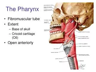
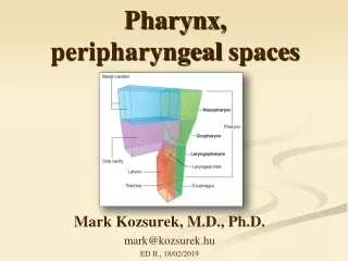
PHARYNX. Pharynx. DEFINATION. It is a muscular tube that extends from the base of the skull to the 6 th cervical vertebra. It is continuous below with the esophagus. It is funnel shaped with an upper expanded end and a narrow lower end. SUBDIVISIONS.

E N D
PHARYNX Pharynx Prof .Saeed Makarem
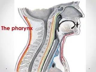
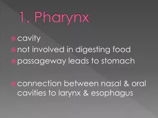
DEFINATION It is a muscular tube that extends from the base of the skull to the 6th cervical vertebra. It is continuous below with the esophagus. It is funnel shaped with an upper expanded end and a narrow lower end Prof. Saeed Makarem
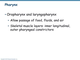
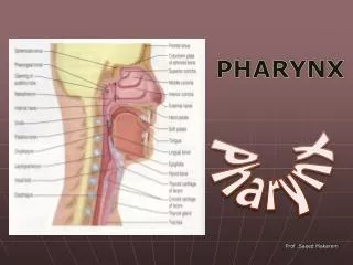
SUBDIVISIONS It lies behind the nose, mouth and larynx. It communicates with each of them. It is divided into 3 parts: Nasopharynx. Oropharynx. Laryngopharynx. So, Its anterior wall is deficient where it is related to nose, mouth & larynx. Prof. Saeed Makarem
RELATIONS Posteriorly: It lies infront of the prevertebral muscles and fascia. Laterally : It is related from above downward to: 1-Auditory tube. 2-Styloid process and its muscles. 3-Carotid sheath and its contents. 4-Thyroid gland Prof .Saeed Makarem
STRUCTURE OF THE WALL From inwards outwards 1-Mucous membrane: It is continuous with that of the Nose, Oral cavity, Auditory tube, Larynx ,esophagus. 2- Pharyngobasilar fascia. 3- Muscles. 4- Buccopharyngeal fascia The 2 layers of Fascia: It is thickened internally to form the Pharyngobasilar fascia and externally to form Buccopharyngeal
MUSCLES OF PHARYNX Prof.Saeed Makarem
MUSCLES Constrictors: 1- Superior. 2- Middle 3- Inferior. Prof.Saeed Makarem
The three constrictors muscles overlap each others like segment of an antenna. Each one fans out from its anterior attachment and passes posteriorly around the pharynx horizontally. Prof.Saeed Makarem
CONSTRICTORS Each muscle joins its fellow of the opposite side in a fibrous midline Pharyngealraphe. The raphe extends from the pharyngeal tubercle of the occipital bone to the esophagus. The constrictors propel the bolus of food down into the esophagus. Prof. Saeed Makarem
CONSTRICTORS The lowest fibers of the inferior constrictor forms the Cricopharyngeusmuscle. It has a sphincteric action on the lower end of the pharynx and prevents the entry of the air into the esophagus in between swallowing. Prof.Saeed Makarem
LONGITUDINAL MUSCLES 1-Stylopharyngeus. 2-Salpingeopharyngeus. 3-Palatopharyngeus. They are attached to the styloid process . Auditory tube. Soft palate. They elevate the larynx and pharynx and pulls the palatopharyngeal arch medially during swallowing Prof .Saeed Makarem
MOTOR INNERVATION • All muscles of the pharyngeal are supplied by the pharyngeal plexus Except the Stylopharyngeuswhich is supplied by the glossopharyngealnerve. Prof .Saeed Makarem
SENSORY INNERVATION Nasopharynx: 5-Maxillary nerve. Oropharynx: 9-Glossopharyngeal Laryngopharynx: 10-Vagus nerve Prof.Saeed Makarem
NASOPHARYNX It has a respiratory function. It extends from skull base to the soft palate. Boundaries: Roof: Basilar part of occipital bone. Body of sphenoid. A collection of lymphoid tissue lies in the submucosa at the junction of roof & posterior wall. it is called pharyngeal tonsil Prof.Saeed Makarem
NASOPHARYNX Floor : Sloping upper surfaceof the soft palate. Posterior wall: Anterior arch of atlas. Pharyngeal isthmus: It is a gap in the floor between the free end of the soft palate and the posterior wall of the pharynx. Prof. Saeed Makarem
NASOPHARYNX LATERAL WALL: it has: 1. Pharyngeal opening of the auditory tube. 2.Tubal elevation: The elevated posterior margin of the tube. 3-Pharyngeal recess: It is a slit like depression behind the tubal elevation. Prof.Saeed Makarem
NASOPHARYNX 4.Salpingo-pharyngeal fold of mucous membrane produced by the salpingopharyngeal muscle. 5.Tubal tonsil It is a collection of lymphoid tissue behind the opening of the auditory tube. Prof.Saeed Makarem
ADENOID Repeated infection of the pharyngeal tonsils leads to its hypertrophy Now it is called adenoids. It causes obstruction of the posterior nares. The patient breathes through his mouth and snores. It can cause deafness and recurrent otitis media due to its close relation to the auditory tube. Prof . Saeed Makarem
OROPHARYNX It has a digestive function. It lies behind the mouth cavity and extends from the soft palate to the upper border of the epiglottis. Roof : It is the under surface of the soft palate and the pharyngeal isthmus. Prof. Saeed Makarem
OROPHARYNX Floor : It is the posterior one third of the tongue and the interval between the tongue and the anterior surface of the epiglottis. Lingual tonsil : A collection of lymphoid tissue on the posterior third of the tongue. Prof .Saeed Makarem
OROPHARYNX The tongue is connected to the epiglottis by one median and two lateral glossoepiglotticfolds. VALLECULA : It is a depression on each side of the of the median glossoepiglottic fold. Prof.Saeed Makarem
OROPHARYNX Anterior wall : It opens into the mouth cavity through the oropharyngeal isthmus. Posterior wall : Body of 2nd cervical vertebra and the upper part of the body of the 3rd vertebra. Prof.Saeed Makarem
OROPHARYNX Lateral wall : Palatoglossal arch It is a mucous fold overlying the palatoglossal muscle. Palatopharyngealarch It is a mucous fold behind the palatoglossal arch. • The interval between the two palatopharyngeal arches is the • Oropharyngeal isthmus Prof .Saeed Makarem
PALATINE TONSIL It occupies the (Tonsillar Bed). Which is the interval between the palatoglossal & palatopharyngeal Prof.Saeed Makarem
PALATINE TONSIL Structure : It is an oval body of lymphoid tissue. It has an upper and lower poles. Its deep surface is attached to a fibrous capsule. Prof.Saeed Makarem
PALATINE TONSIL RELATIONS : Lateral: superior constrictor muscle and fascia. The external palatine vein lies within this fascia. The facial artery is more lateral. Medial : Oropharyngeal isthmus. Prof.Saeed Makarem
PALATINE TONSIL Superior: Soft palate. Here the tonsil is continuous with the lymphoid tissue on its undersurface . Inferior : Posterior third of tongue. It is continuous with the lingual tonsil. Prof.Saeed Makarem
PALATINE TONSIL Arterial Supply : Tonsillar branch of facial artery. Tonsillar branch of ascending palatine. The arteries enter its deep surface. Venous Drainage: The veins pierces the superior constrictor and join the external palatine, the pharyngeal and or the facial veins vein. .
PALATINE TONSIL Lymph Drainage: Jugulodigastric nodes (which lies below and behind the angle of the mandible). Prof .Saeed Makarem
TONSILITIS The tonsils have their maximum size in childhood. They are a common site of infection. This is manifested by sore throat ,pyrexia and tender enlarged jugulodigastric lymph nodes. After tonsillectomy ,the external palatine vein may be the source of postoperative bleeding. Prof.Saeed Makarem
PERITONSILAR ABSCESS QUINSY It is causedby the spread of infection from the palatine tonsil to the loose connective tissue outside the capsule. Prof. Saeed Makarem
LARYNGOPHARYNX It extends from the upper border of the epiglottis to the lower border of the cricoid cartilage (C6). Anterior wall: Inlet of the larynx. Posterior wall: Bodies of 3rd , 4th ,5th and 6th cervical vertebrae. Prof. Saeed Makarem
LARYNGOPHARYNX Lateral wall: Thyroid cartilage. Thyrohyoid membrane. Prof .Saeed Makarem
PIRIFORM FOSSA It is a small depression on either side of the laryngeal inlet. It is separated from the laryngeal inlet by the Aryepiglottic fold. Prof .Saeed Makarem
PIRIFORM FOSSA It is bounded Laterally by the thyroid cartilage and thyrohyoid membrane. Branches of the internal laryngeal and recurrent Laryngeal nerves lie deep to its mucous membrane. Prof. Saeed Makarem
PIRIFORM FOSSA They are liable to be injured when a foreign body ( bony fish) is lodged in the fossa. A foreign body in the fossa causes the patient to gag violently, and needs a physician’s assistance Prof .Saeed Makarem