Download
1 / 69
910 likes | 1.89k Views
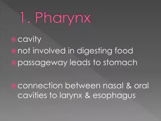
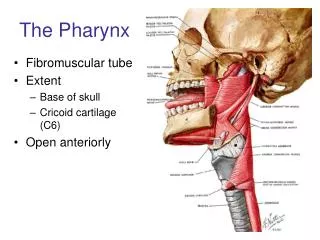
PHARYNX. Introduction. Musculomembranous tube, 12 cms Basiocciput to cricoid lower border (C6) Relation Superior basiocciput Inferior oesophagus Posterior prevertebral fascia with muscles Lateral digastric and carotid triangle Anterior

E N D
Introduction Musculomembranous tube, 12 cms Basiocciput to cricoid lower border (C6) Relation • Superior • basiocciput • Inferior • oesophagus • Posterior • prevertebral fascia with muscles • Lateral • digastric and carotid triangle • Anterior • Wall deficient, communicate with naso, oro, & laryngopharynx • Pharyngeal isthmus : junction with NP & OP • Oropharyngeal isthmus : junction with OP & oral cavity
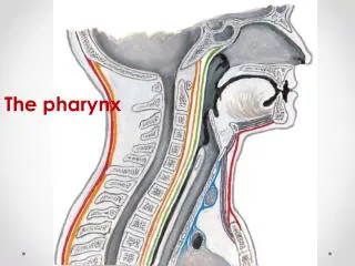
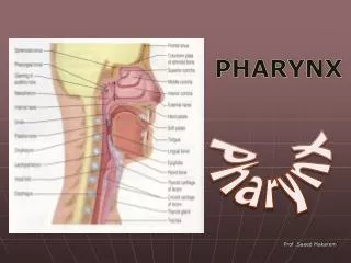
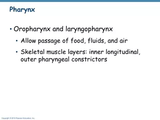
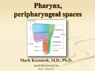
Parts of pharynx Nasopharynx Oropharynx Laryngopharynx
Oropharyngeal isthmus Oropharynx behind palatoglossal arch
Median Section: Pharyngeal isthmus Pharyngeal isthmus
Laryngeal msls Posterior view : wall of pharynx dissected
Nasopharynx Base of skull to soft palate Features • Opening of ET : lateral wall • Tubal elevation(upper post to ET): cartilagenous part of ET • Folds : (post to tubal elevation) • Salpingopharyngeal fold : salpingopharyngeus • Salphingopalatine fold: levator palati • Pharyngeal recess (fossa of Rosenmuller) • Behind opening of AT • Misguided catheter may perforate ICA • Pharyngeal tonsil • Lymphatic tissue at roof (adenoids) • Tubal tonsil • Lymphatic tissue at region of TE
Nasopharynx Parts of pharynx
Oropharynx SP lower border to epiglottis superior border • Palatoglossal arch • Palatopharyngeal arch • Palatine tonsil Laryngopharynx Epiglottis sup border to cricoid cart inf border • Ant : cavity of larynx • Aryepiglottic fold • Piriform recess • lateral to folds and behind lateral glossoepiglottic fold
Coats of pharynx • Mucous membrane • NP : pseudostratified columnar • LP/OP : stra. squamous ciliated columnar • Fibrous layer (pharyngobasilar fascia) • Thickened upper part : support NP, keep patent • Median raphe : basiocciput to cricoid • Muscular layer • Inner longitudinal • Outer circular • Fascial layer (buccopharyngeal fascia) • Formed of epimysium of constrictors
Muscles of pharynx Inner longitudinal • Stylopharyngeus • Salpingopharyngeus • Palatopharyngeus • All insertions to post border of thyroid cart • Salpingopharyngeus blends with palatopharyngeus • Action : pull pharynx during 3rd stage of deglutition
Stylopharyngeus • From base of styloid pr to post border of thyroid • Enter b/t SC & MC Salpingopharyngeus • From AT cart, and merges with PP, inserts to post border of Thyroid Palatopharyngeus • From palate to post border of thyroid & side of pharynx • Form Passavant’s ridge on post pharyngeal wall
Relation of SP, PP & Salpingopharyngeus to constrictors Form Passavant’s ridge
SP PP SP
Muscles of pharynx Outer circular - Constrictors • constrict in sequential manner to push bolus down • Types • Superior constrictor • Middle constrictor • Inferior constrictor
Superior constrictor Origin • Lower medial pterygoid plate • Pterygoid hamulus • Pterygomandibular raphe • Mylohoid line posterior part • Tongue laterally in the posterior part Fibres insertion • Upper – Pharyngeal tubercle, medial raphe, • Lower – medial raphe till cricoid cart • Middle - horizontal
Shows ligaments, raphe & sites of attachments of muscles Thyrohyoid membrane
Muscles of Pharynx Stylopharyngeus entering b/t SC and MC
Middle constrictor Overlaps sup constrictor Origin • Styloid lig • Lesser horn hyoid bone • Greater horn upper border Fibres • Upper – horizontal, enclose Sup constrictor • Lower – run obliquely down to medial raphe
Inferior constrictor Two parts : Thyropharyngeus • Origin – thyroid oblique line, tendinous arch • Fibres • Upper – run upwards • Lower – run horizontal to median raphe • Propulsive action Cricopharyngeus • Origin – cricoid cart lat surf • Fibres – • run horizontal uninterruptedly from one side to other (does not insert to median raphe) • Sphincteric action at lower extent of pharynx • Continuous with circular mls of oesophagus
ligaments, raphe & sites of attachments of muscles Thyrohyoid membrane
Pterygomandibular raphe Structures passing through gaps between constrictors
Gaps between constrictors • Space of Morgagni • Space b/t SC & MC • Space b/t MC & IC • Space b/t IC & oesophagus
Gaps between constrictors Killian’s dehiscence Weak muscular wall b/t thyropharngeus & cricopharyngeus Pharyngeal diverticulum Space of Morgagni • Covered by Pharyngobasilar fascia • LP, TP, Auditory tube • Ascending palatine art Space b/t SC & MC • Stylopharyngeus • Nerve : IX, Lingual Space b/t MC & IC • Covered by Thyroglossal membrane • IL nerve • Sup laryngeal Vs Space b/t IC & oesophagus • RL nerve • Inf laryngeal vessel
Nerve supply Pharyngeal plexus – (supply pharynx except SP) • Over MC, formed by : • X nerve pharyngeal br – motor and sensory • IX nerve pharyngeal br (purely afferent i.e. sensory) • Superior cervical ganglion for sympathetic supply motor : • By pharyngeal plexusexceptstylopharyngeus (IX cranial nerve) • Cr part of XI nerve, travel along X nerve • Cricopharyngeus : RLN or ELN (NA – Nucleus ambiguus is a motor nucleus for IX, X and XI nerves) Sensory: • NP : Maxillary nerve through pterygopalatine ganglion • OP : IX nerve • LP : X nerve br Sympathetic supply : vasomotor From pharyngeal plexus components