Download
1 / 37
520 likes | 1.54k Views
The pharynx. The pharynx. Anatomy of The pharynx Site. Seen from behind. Midline of the neck From skull base to esophagus In front of upper 6 Cervical vertebra. Behind : The Nose The Mouth The larynx. Irregular Fibromuscular tube lined by mucous membrane Length: 15 cm. Shape.

E N D
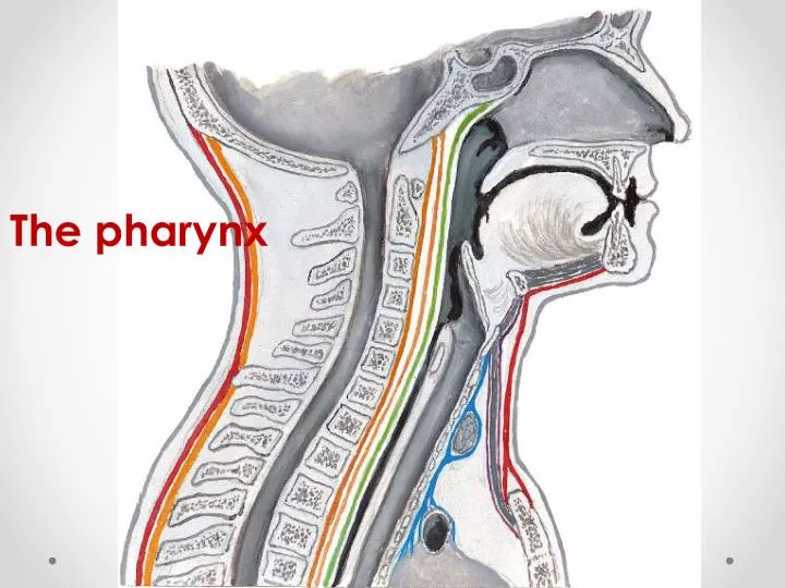
The pharynx The pharynx
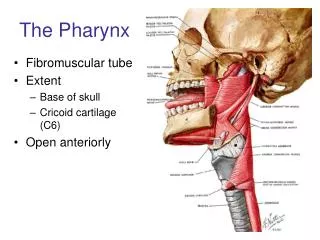
Anatomy of The pharynxSite Seen from behind Midline of the neck From skull base to esophagus In front of upper 6 Cervicalvertebra Behind : The Nose The Mouth The larynx
Irregular Fibromuscular tube lined by mucous membrane Length: 15 cm Shape
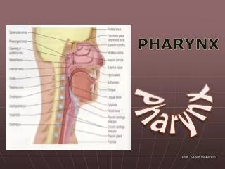
Nasopharynx Oropharynx Laryngopharynx (Hypopharynx) the pharynxCompartments Seen from behind
Seen from lateral • Nasopharynx • Oropharynx • Laryngopharynx (Hypopharynx)
Muscles of the pharynx • Circular muscles: Superior, middle and inferior constrictor muscles. • Longitudinal muscles:stylopharyngeus, palatopharyngeus and salpingopharyngeus • All muscles of the pharynx innerveated by cranial root of accessory nerve except stylopharyngeus which is supplied by glossopharyngeus nerve
Three constrictor stylopharyngeus cricopharyngeus
-Behind the nasal cavity -Extends from skull Base superiorly to the soft palate inferiorly Communicates inferiorly with the oropharynx through the pharyngeal isthmus The nasopharyngeal tonsil lies in the roof The pharyngeal opening of ET lies in the lateral wall Nasopharynx
Nasopharynx (important landmarks) • The pharyngeal isthmus • Opening of auditory tube and tubal elevation • The salpingopharyngeal fold
Behind the oral cavity (in front of 2nd&3rd Cervical vertebra) From the soft palate superiorly to tip of epiglottis inferiorly Communicates: Anteriorly with the oral cavity Superiorly with the nasopharynx Inferiorly with the laryngopharynx The paatine tonsils lie laterally between the anterior and posterior pilars Oropharynx
The floor • Median epiglottis fold vallecula lateral epiglottis fold
Behind the Larynx(in front of 3rd to 6th Cervical vertebra) From the tip of epiglottis superiorly to the lower border of cricoid cartilage Inferiorly Communicates: Anteriorly with the Larynx Superiorly with the oropharynx Inferiorly with the esophagus laryngopharynx
The hypopharynx does not only lie behind the larynx BUT also Projects laterally on each side of the larynx So it is formed of : Postcricoid region ( behind the larynx) Two pyriform fossa (on each side of the larynx Seen from behind Cross section
What is Waldeyer’s ring? The lymphoid tissue in the pharyngeal aponeurosis aggregates in some areas forming tonsils: 1-onenasopharyngeal tonsil 2- twopalatine tonsils 3- twolingualtonsils 4-twotubaltonsils
Nerve Supply Motor ---►XIExcept : Stylopharyngeus--►IX Sensory--► • Nasopharynx: V • Oropharynx: IX • Laryngopharynx: X Autonomic: • sympathetic: SCG • Parasympathetic: through VII
Palatine tonsils • Defintion • Site • Covering • Variability in size • Blood supply • Lymph drainage
The anterior pillar formed by palatoglossus muscle The posterior pillar formed By palatopharyngeus m The tonsils lie between the Two pillars