Download
1 / 36
370 likes | 381 Views
INTRODUCTION M SE 206 - Materials Characterization I. Course Syllabus Aim:

E N D
INTRODUCTION MSE 206-Materials Characterization I
Course Syllabus Aim: The aim of this course is to familiarize the students with the fundamentals of microstructural characterization, basics of geometric optics and light microscopy. Both theoretically and practically, the students will have the chance to learn how to work on an optical microscope and determine typical microstructures of ferrous as well as nonferrous metallic materials.
Textbook • Textbook: There is no recommended specific textbook in metallography in the English language. For this reason the students are asked to attend the lectures regularly and take notes. Relevant class material will be posted onwww.mse206.cankaya.edu.tr
Laboratory • In the metallography lab, each student is assigned to a microscope and she/he will be held responsible to look after it well. Lab schedule is handed out in the class. Pop quizzes will be given in each lab hour. In addition to this, students are required to complete lab reports each week. • Term Project: Each student will be given a metallic sample of unknown composition and microstructure. Using the necessary characterization tools available in our department the student will try to determine the alloy composition, microstructure, hardness etc. of the unknown sample. Based on the obtained information the student will comment on the processing as well as possible heat treatment history of the sample and typical applications of the alloy with the determined properties. The term projects are due the last day of classes.
Attendance&Grading Attendance 70% attendance of all lecture hours and 80% attendance of all laboratory hours is required by the university’s regulations. Absence from a quiz, lab. or an examination will result in zero grade. Grading Policy
Weekly Lab Topics • Metallographic Specimen Preperation • Optical Microscopes • Macro-examination, Inclusions andSolidification Structures • Equilubrium cooled Fe-C Structures • Isothermal Transformation Fe-C Structures • Non-Equilubrium Structures • Cast Irons • Plastically Deformed and Surface TreatedStructures • Nonferrous Metallography • Scanning Electron Microscopy • Transmission Electron Microscopy • Quantitative Metallography
Materials Optimization Loop Material science is the investigation of the relationship among processing, structure, properties, and performance of materials.
Structure 1) Sub atomic – electrons and nuclei (protons and neutrons) 2) Atomic – organization of atoms or molecules 3) Microscopic – groups of atoms that are normally agglomerated together 4)Macroscopic – viewable with the un-aided eye 1 Ao = 10-10 m 1 nm = 10-9 m 1 m = 10-6 m 1 mm = 10-3 m 1 cm = 10-2 m
Property - A material trait expressed in terms of the measured response to a specific imposed stimulus • Hardness • Strength • Ductility, etc. • Resistivity • Conductivity • Mag. permeability • Ther. conductivity • Heat capacity • Index of refraction • Reflectivity + Deteriorative Properties: Relate to the chemical reactivity of materials
Why Materials Characterization is Necessary ? Properties (mechanical, electrical&magnetic, optical, thermal) of materials depend on internal structure (in microscopic and atomic level ; bonding, crystal structure and composition) • Characterization is needed to identify the material and predict material properties, as well as processing conditions • Some characterization techniques are qualitative,such as providing an image of a surface, others yield quantitative informationsuch as the relative concentrations of atoms that comprise the material. Qualitative – what is present? Quantitative – how much is present?
Categories of Materials Characterization Techniques 1) Image 2) Surface 3) Structural 4) Organic 5) Elemental
1)Image Analysis • Optical microscopy • Confocal microscopy • SEM/EDX – Scanning Electron Microscopy with Energy Dispersive X-Ray detector • SPM – Scanning Probe Microscopy • AFM – Atomic Force Microscopy • TEM – Transmission Electron Microscopy
1)Image Analysis p-n ZnO nanowires by SEM
Surface Analysis • AES – Auger Electron Spectroscopy • XPS – X-Ray Photoelectron Spectroscopy • TOF-SSIMS – Time of Flight Static Secondary Ion Mass Spectroscopy • LEED – Low Energy Electron Diffraction
Surface Analysis XPS – X-Ray Photoelectron Spectroscopy: gives information from 1-10 nm depth. XPS results of Titanium surface Sample Binding Energies (eV)
Structural Analysis • XRD – X-Ray Diffraction • XAX/EXAFS - X-ray Absorption Spectroscopy and Extended X-Ray Absorption Fine Structure • Raman spectroscopy • TEM – Transmission Electron Microscopy • EELS – Electron Energy Loss Spectroscopy (typically combined with TEM)
Organic Analysis • FTIR – Fourier Transform Infrared Spectroscopy • GC/MS – Gas Chromatography with Mass Spectroscopy (detector) • HPLC – High Performance Liquid Chromatography • Raman spectroscopy (structural organic)
Elemental Analysis • ICP – Inductively Coupled Plasma • XRF – X-Ray Fluorescence • PIXE - Particle-Induced X-ray Emission • Optical atomic spectroscopy • CHN (Carbon / Hydrogen / Nitrogen)
Scope of ‘’Materials Characterization I’’ Course Some Image Characterization Techniques: 1-Light (optical) microscopy (LM) or (OM) 2-Scanning electron microscopy (SEM) Energy dispersive X-ray spectroscopy (EDS) 3-Transmission electron microscopy (TEM)
Electron Microscopes vs. Optical Microscope • Optical Microscope uses visible light (photons, 0.4-0.7 m wavelength). Contrasts in the image produced result from differences in reflectivity of the various regions of the microstructure. • Transmission Electron Microscope (TEM) uses a wide beam of electrons passing through a thin sliced specimen to form an image. • Scanning Electron Microscope (SEM) uses focused beam of electrons scanning over the surface of thick or thin specimens.. Images are produced one spot at a time in a grid-like raster pattern. (will be discussed in a later lecture)
Electron Microscopes vs. Optical Microscope Transmission Electron Microscope, TEM Optical Microscope Scanning Electron Microscope, SEM
Resolution Scale of Different Methods XRD,TEM,STMSEM OM Valve Turbo charge Grain I Grain II Atomic atomic
IMAGES: OM vs SEM Optical Microscope image of a polycrystalline material BaTiO3 50m Scanning Electron Microscope image of a polycrystalline BaTiO3 5m Optical microscopy provides 2-dimensional image whereas SEM is able to show 3-dimensional features
Identification of Fracture Mode by SEM Pores Cracks Cracks Grain boundary 4m 20m Intergranular fracture Intragranular fracture
SEM Images Pollen Butterfly Tongue Fire ant head Spider
SEM Images flue virus Snow crystals
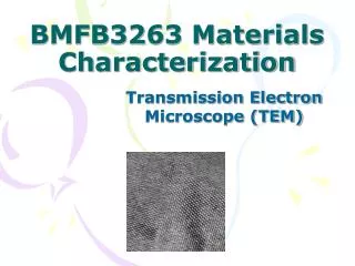
Transmission Electron Microscopy Schematic view of carbon nanotubes TEM image of carbon nanotubes
Transmission Electron Microscopy TEM imaging of graphene
Transmission Electron Microscopy Dislocations TEM image of deformed 316 stainless steel showing dislocations and dipoles on (111) planes.
Transmission Electron Microscopy Close view of TEM images of some nano-catalysts.
Homework-I Write a short report about; Similarities and differences between optical (light), Scanning electron microscope (SEM) and Transmission Electron Microscopes (TEM)