Download
1 / 1
10 likes | 141 Views
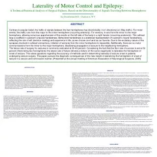
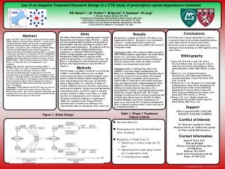
Site. Pt. #. location. AM. AR-i. AR-s. AR-r. FM. FE. FR-i. FR-s. FR-r. A. 1. AG. 0. X. 0. 0. NT. 0. NT. NT. NT. B. 1. IPL. 0. 0. X. 0. NT. 0. NT. NT. NT. C. 2. STG. 0. 0. 0. 0. 0. 0. 0. X. 0. D. 3. STG. †. †. †. †. X. †. 0. 0. 0. E. 5.

E N D
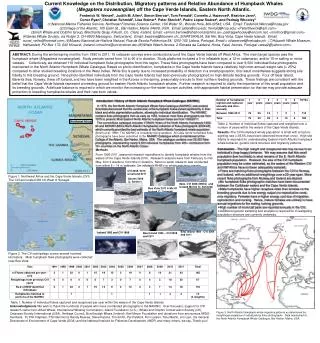
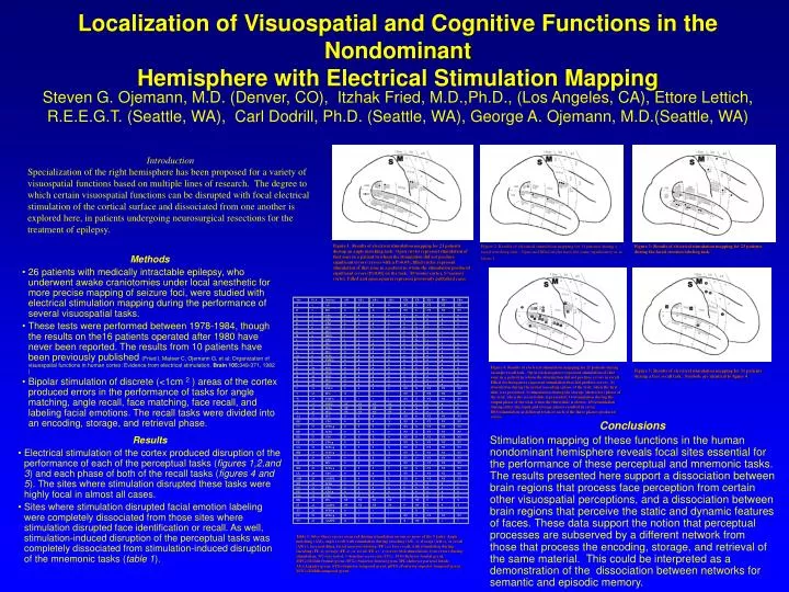
Site Pt. # location AM AR-i AR-s AR-r FM FE FR-i FR-s FR-r A 1 AG 0 X 0 0 NT 0 NT NT NT B 1 IPL 0 0 X 0 NT 0 NT NT NT C 2 STG 0 0 0 0 0 0 0 X 0 D 3 STG † † † † X † 0 0 0 E 5 STG 0 X 0 0 0 0 0 0 0 F 6 IFG † † † † 0 X † † † G 6 IFG † † † † 0 X † † † H 6 MFG † † † † 0 X † † † I 6 STG † † † † 0 X † † † J 7 IPL X 0 0 0 0 0 0 0 0 K 7 IFG X 0 0 0 X 0 0 0 0 L 8 IFG 0 0 0 X 0 0 0 0 0 M 8 MTG 0 0 0 0 0 X 0 0 0 N 8 PMTG 0 0 0 0 0 X 0 0 0 O 9 STG 0 0 0 0 0 0 0 0 X P 10 SFG 0 0 0 0 0 X 0 0 0 Q 10 MFG 0 0 0 0 0 0 X 0 X R 10 SFG 0 0 0 0 0 0 0 X 0 S 10 IFG 0 0 0 0 X 0 0 0 0 T 10 IFG 0 0 0 0 0 0 X 0 0 U 12 PSTG 0 0 0 0 NT X NT NT NT V 13 IFG X † † † NT 0 NT NT NT W 13 PMTG 0 † † † NT X NT NT NT X 15 PSTG NT NT NT NT X 0 0 0 0 Y 15 MTG NT NT NT NT 0 X 0 0 0 Z 16 IFG NT NT NT NT X 0 0 0 0 AA 17 IFG 0 0 0 X NT 0 NT NT NT BB 17 STG 0 0 0 X NT 0 NT NT NT CC 17 MTG-p 0 0 0 0 NT X NT NT NT DD 17 MTG 0 0 0 0 NT X NT NT NT EE 17 STG 0 X X 0 NT 0 NT NT NT FF 17 STG-p 0 0 0 0 NT X NT NT NT GG 18 MTG-p 0 0 0 X NT 0 NT NT NT HH 19 MTG-p 0 X X X NT 0 NT NT NT II 19 STG 0 X 0 0 NT 0 NT NT NT JJ 19 IPL 0 0 X 0 NT 0 NT NT NT KK 20 MTG-p 0 0 0 0 NT X NT NT NT LL 20 STG 0 0 0 X NT 0 NT NT NT MM 20 AG/IPL 0 0 X 0 NT 0 NT NT NT NN 21 MTG 0 0 0 0 0 X 0 0 0 OO 21 STG 0 0 0 X 0 0 0 0 0 PP 21 STG-p 0 X 0 0 0 0 0 0 0 QQ 21 STG 0 X 0 0 0 0 0 0 0 RR 22 IPL NT NT NT NT 0 NT 0 0 X SS 22 AG/IPL NT NT NT NT 0 NT 0 0 X TT 23 MTG-p 0 † † † * X * * * UU 25 MTG-p X † † † NT 0 NT NT NT VV 26 AG/IPL X † † † X NT † † † Localization of Visuospatial and Cognitive Functions in the NondominantHemisphere with Electrical Stimulation Mapping Steven G. Ojemann, M.D. (Denver, CO), Itzhak Fried, M.D.,Ph.D., (Los Angeles, CA), Ettore Lettich, R.E.E.G.T. (Seattle, WA), Carl Dodrill, Ph.D. (Seattle, WA), George A. Ojemann, M.D.(Seattle, WA) Introduction Specialization of the right hemisphere has been proposed for a variety of visuospatial functions based on multiple lines of research. The degree to which certain visuospatial functions can be disrupted with focal electrical stimulation of the cortical surface and dissociated from one another is explored here, in patients undergoing neurosurgical resections for the treatment of epilepsy. Figure 1: Results of electrical stimulation mapping for 21 patients during an angle matching task. Open circles represent stimulation of that zone in a patient in whom the stimulation did not produce significant errors (errors with a P>0.05), filled circles represent stimulation of that zone in a patient in whom the stimulation produced significant errors (P≤0.05) on the task. M=motor cortex, S=sensory cortex. Filled and open squares represent previously published cases. Figure 2: Results of electrical stimulation mapping for 11 patients during a facial matching task. Open and filled circles have the same significance as in figure 1. Figure 3: Results of electrical stimulation mapping for 23 patients during the facial emotion labeling task. • Methods • 26 patients with medically intractable epilepsy, who underwent awake craniotomies under local anesthetic for more precise mapping of seizure foci, were studied with electrical stimulation mapping during the performance of several visuospatial tasks. • These tests were performed between 1978-1984, though the results on the16 patients operated after 1980 have never been reported. The results from 10 patients have been previously published (Fried I, Mateer C, Ojemann G, et al: Organization of visuospatial functions in human cortex: Evidence from electrical stimulation. Brain 105:349-371, 1982 ) • Bipolar stimulation of discrete (<1cm 2 ) areas of the cortex produced errors in the performance of tasks for angle matching, angle recall, face matching, face recall, and labeling facial emotions. The recall tasks were divided into an encoding, storage, and retrieval phase. Figure 4: Results of electrical stimulation mapping for 15 patients during an angle recall task. Open circles/squares represent stimulation of that zone in a patient in whom the stimulation did not produce errors in recall. Filled circles/squares represent stimulation that did produce errors. I= stimulation during the initial (encoding) phase of the trial, when the first slide was presented, S=stimulation during the storage (distractor) phase of the trial, when the second slide is presented; O=stimulation during the output phase of the trial, when the third slide is shown. IS=stimulation during either the input and storage phases resulted in error, ISO=stimulation at different trials of each of the three phases produced errors Figure 5: Results of electrical stimulation mapping for 11 patients during a face recall task. Symbols are identical to figure 4. Conclusions Stimulation mapping of these functions in the human nondominant hemisphere reveals focal sites essential for the performance of these perceptual and mnemonic tasks. The results presented here support a dissociation between brain regions that process face perception from certain other visuospatial perceptions, and a dissociation between brain regions that perceive the static and dynamic features of faces. These data support the notion that perceptual processes are subserved by a different network from those that process the encoding, storage, and retrieval of the same material. This could be interpreted as a demonstration of the dissociation between networks for semantic and episodic memory. • Results • Electrical stimulation of the cortex produced disruption of the performance of each of the perceptual tasks (figures 1,2,and 3) and each phase of both of the recall tasks (figures 4 and 5). The sites where stimulation disrupted these tasks were highly focal in almost all cases. • Sites where stimulation disrupted facial emotion labeling were completely dissociated from those sites where stimulation disrupted face identification or recall. As well, stimulation-induced disruption of the perceptual tasks was completely dissociated from stimulation-induced disruption of the mnemonic tasks (table 1). Table 1: Sites where errors occurred during stimulation on one or more of the 5 tasks: Angle matching (AM), angle recall with stimulation during encoding (AR –i), storage (AR-s), or recall (AR-r), face matching, facial emotion labeling (FE), or face recall, with stimulation during encoding (FE-i), storage (FE-s), or recall (FE-r). x=errors with stimulation. 0=no errors during stimulation, NT=not tested, †=baseline error rate>25%. IFG=Inferior frontal gyrus, MFG=Middle frontal gyrus, SFG= Superior frontal gyrus, IPL=Inferior parietal lobule, AG=Angular gyrus, STG=Superior temporal gyrus, pSTG=Posterior superior temporal gyrus. MTG=Middle temporal gyrus.