Download
1 / 2
20 likes | 152 Views
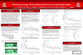
Laterality of Motor Control and Epilepsy: A Technical/Statistical Analysis of Surgical Failures, Based on the Directionality of Signals Traveling Between Hemispheres Iraj Derakhshan M.D., Charleston, W.V. ABSTRACT

E N D
Laterality of Motor Control and Epilepsy: A Technical/Statistical Analysis of Surgical Failures, Based on the Directionality of Signals Traveling Between Hemispheres Iraj Derakhshan M.D., Charleston, W.V. ABSTRACT Contrary to popular belief, the traffic of signals between the two hemispheres has directionality; it is1-directional (a1-Way traffic). For motor events, the traffic runs from the major to the minor hemisphere (occurring anteriorly). For sensory, it runs from the minor to the major hemisphere, allowing conscious apprehension of the events on the left side of the body in a right hander (occurring posteriorly). This callosal loop is codified in a person’s (neural) handedness. Behavioral handedness is a statistical representation of a person’s neural handedness, reflecting the role of will (decision making) and experience in life, as we choose one hand as our favorite. Due to the excitatory nature of the synapses involved in callosal connections, initiation of seizures from the minor hemisphere is impossible. Additionally, there are no motor communications from the minor to the major hemisphere, disallowing propagation of seizure to the neighboring hemisphere. The failure rate of surgery for seizures is commonly estimated at 35-40 percent. Considering the fact that the floor rate of success is set at 50 percent (there being two hemispheres) the above rate of failure denotes a failure (of the same magnitude) to lateralize the hemisphere of onset of seizure. This raises questions regarding the accuracy of methods used in determining laterality of seizure onset in patients undergoing seizure surgery. This paper pursues the diagnostic consequences of the new insight in lateralizing the hemisphere of onset of seizure in a secure and noninvasive manner. [Presented at the annual meeting of American Association of Neurological Surgeons, 2005] INTRODUCTION RESULTS Since there is no motor communication from the right to the left hemisphere in right handers and all callosal connections between hemispheres are excitatory, 1, 2 seizures may not initiate in or propagate to the left from the right hemisphere (in right hander). Note: Seizures arising from raised intracranial pressure due to expanding lesions in the minor hemisphere may not count against the above generalization as the major hemisphere remains susceptible to such events in those circumstances.It is to be remembered that papilledema, indicating raised intracranial pressure, occurred in 36 percent of patients with temporal lobe tumors (Bingley, see below). The dichotomous nature of the command structure reduces the status of earlier considerations, suggesting existence of bi-hemispheric “continuum” for speech and other tasks, to ones’ arising from lack of temporal resolution of the tests involved including routine EEGs. It has been shown that routine EEG does not have the resolution to determine temporal precedence of seizure activity in the major/lead hemisphere. 2, 5 Nor can the commonly used criteria of amplitude of seizure activity be relied upon for this purpose because of its susceptibility to a variety of factors. Some of these confounding factors are known to clinicians familiar with electrodiagnosis of carpal tunnel syndrome, including the amount pressure applied to the recording site. 2, 6 As an example of confusion caused by amplitude criteria one may cite the case of a neural left hander in whom the potentials arising from lead hemisphere (right) were of lower amplitude compared to those recorded over the minor (left) hemisphere. 7 In another study by Blume on hemispheric epilepsy involving 13 selected cases, the amplitude of seizure potentials were higher on the side of the minor hemisphere more often (8/13) than they were on the side of the major hemisphere.8 Documenting temporal precedence of seizure activity in one hemisphere compared to the other is feasible and has been satisfactorily employed in determining laterality of the major hemisphere. 2, 5, 7, 9, 10 Such determinations will resolve the questions surrounding the issue of bilateral “independent” seizure foci and the related subject dealing with “shifting of sides” of seizure activity. 2, 11, 12 That behavioral handedness may not be relied upon in affirming the laterality of motor control (except in its statistical sense, see above) has been known to classical neurology since the time of Bramwell’s, who first described a case of crossed aphasia (1899). More recent examples of similar occurrences are on record using modern techniques. 13 In this respect, no less that 15 percent of the public is incongruent in their neural and behavioral handedness, the majority of whom among the left handers. It is the presence of this significant minority (one in five persons) that lies at the heart of the quest for ascertaining a surgical candidate’s neural handedness; i.e. the laterality of the hand closer to the command center by a callosum-width. A fundamental contribution of 1-way callosal traffic circuitry was to restore the “constant conjunction” of (neural) handedness and speechedness by demonstrating that the command center issues all commands (including speech) regardless of the laterality of the effectors carrying out the same. The newly discovered vectorial relationship between the hemispheres provides for error-proof determination of laterality of command center if the patient is able to perform tasks involving push of a button or turning of the eyes to the sides (reaction time determination). The sensory counterpart of the loop may also be explored by comparing the limbs for sensory evoked potentials (SEPs), event related desynchronization or metabolic uptake studies. In all of these tests, bilateral signal occurrence upon moving or stimulating a limb indicates the nondominant status of that side of the body. 2, 13, 14 On the other hand, in reaction time studies the hand closer to the major left hemisphere (right, in right handers) will have a shorter reaction time by an IHTT. A notable exception in this regard is seen in sterno-cleido-mastoid muscle where the delay occurs on the right side, due to reversed-laterality of insertion of that muscle to the skull. 15 Further data supporting the existence of an invariable leading hemisphere in driving epilepsy comes from the use of intracarotid injection of phenobarbital to stop status epilepticus or the use of Cardiazol to generate seizure. The available data shows that injecting phenobarbital in the major hemisphere suppresses the ongoing seizure activity in both hemispheres. Injecting phenobarbital or Metrazol in the carotid artery of the minor hemisphere only affects the epileptic process in that hemisphere. 5, 16-20 Significantly, as reported recently, simply recording the seizure at higher speeds was enough to demonstrate the earlier occurrences of seizure activity in the major hemisphere prior to the appearance of the same in the minor (~ 50 milliseconds lag time). 5, 18 In the past, absence of the knowledge concerning dichotomous nature of motor control (laterality of command center, including speech) and reliance on Wada test has resulted in ablation of eloquent cortex in many instances (including the removal of the entire major hemisphere). 2, 5, 18 To Sum: The timing procedure delineated above lateralizes the onset hemisphere of seizures which is always the same as the major hemisphere. It does so based on incontrovertible evidence that the major hemisphere is the seat of initiation of all motor activity including speech. The method is secure and noninvasive. 1, 2 Timing is an essential feature of brain activity. For example, movements are coordinated in time and events are ideally reminisced in order of their occurrence. In this light, coordination and memory are examples of the brain acting as a timing machine in unconscious and conscious realms, respectively. It has been shown that the corpus callosum is involved in both of these activities. Bimanual coordination and the recall of the events occurring on or towards the nondominant side of the body are two commonly occurring examples. Although both of these activities (execution of commands and appreciation of events occurring on/to the left) require the sharing of resources available only in the major hemisphere, moving and sensing the left side of the body are bi-hemispheric events (requiring activation of both hemispheres, see below). 1, 2 It is important to remember that any one may suffer a seizure under appropriate circumstances (for example, physical exhaustion or lack of sleep). What is relevant, therefore, are the factors involved in lowering the seizure threshold at the molecular level and the anatomy governing the spread of seizure from one hemisphere to the other. The following data further substantiates that the anatomy sustaining laterality of motor control (coded in handedness) is the same as that governing laterality of seizure onset. 2 This knowledge allows noninvasive anderror free determination of a person’s laterality of motor control (and seizure onset). METHODS Using the yard-stick of time, it has been established that events on the dominant side of the body (right in right handers) occur sooner than those on the nondominant side by an interval commensurate to the time taken for the signal to reach from major to the minor hemisphere that handles the nondominant side (left in right handers), i.e. the interhemispheric transfer time (IHTT).1-3 This means that the right hemisphere is devoted to events occurring on the left side of the body, carrying out the commands coming from the major hemisphere. Numerous experiments have shown that the reverse holds in regards the sensory signals. This is because signals arising from the left side are first transferred from the right hemisphere to the left before they are appreciated by the person residing there. 1, 2 In this manner the two hemispheres are united into a cohesive brain and the unity of mind/purpose is assured. As in other tasks, the control of language is dichotomously represented, occurring either on the left or the right hemisphere (never both). This laterality is coded in handedness. It has also been established that macular vision is the province of the major hemisphere. 1 The oldest known every-day-life manifestation of the directionality in callosal traffic in the motor arena is a musicological observation named the “melody lead of the right hand in piano players”: Written notes intended for simultaneous performance are delivered ~ 70 milliseconds apart, an interval commensurate with IHTT. Thus, the right hand, playing the melody, is always ahead of the left delivering the harmony. The circuitry delineated above makes it impossible to touch a plane surface (the key board) with both hands at the same time. Ordinary life does not lend itself to recognizing the same callosal-width differential in the sensory arena; however, experimental evidence for the role of callosum in the appraisal of “simultaneity” was documented by Efron in 1963 and has since been confirmed repeatedly. 4 In addition, clinical neurology does not recognize a diaschitic syndrome representing presence of motor communication from the minor to the major hemisphere in lesions affecting the callosum or the minor hemisphere. On the other hand, neurology is replete with examples of such syndromes when the minor hemisphere is cut off from the excitatory influences of the major as a result of callosal injury. These cause motor symptoms on the nondominant side of the body (i.e. a special kind of paresis called apraxia, ipsilateral to the major hemisphere). 1, 2 DISCUSSION Insisting on “bilateralism” of command center or on its ability to move from one side to the other after injury (plasticity, re-organization) is tantamount to rejecting the neuron theory and returning to the reticularism that reigned before the advent of the neuron theory.2, 21Whereas the limitation of the timing technique based on the above-mentioned circuitry hinges on the ability of the subject to perform simple tasks, those of the methods currently in vogue are far more serious; 22 ignoring for now the hazards related to their invasiveness. Thus, in assessing reliability of tests employed for lateralizing the onset/lead hemisphere a useful yard-stick is their liability to assigning bilateralism to motor tasks and the variability of such determinations. Tests that are least susceptible to bilateralism are to be preferred. Incidence of bilateralism in language for the Wada test has varied from 0.5- 65 percent in various reports and those of some of the other tests are given in the tables. 23-26 The question whether the left hemisphere is more likely to engage in epileptic activity has been raised before. 27, 28 Statistically, the key question is whether the previously reported laterality of seizure onset have a distribution similar to that of neural handedness? Although the literature on this issue is relatively meager, substantive critical EEG data points to an onset-asymmetry ratio (as judged by unilateral ictal speech disturbances in right handers) similar to that of neural handedness; i.e. a distribution heavily biased to the left (left 67/ right 33 percent).29, 30 On the other hand, recent reports question the significance or even the utility of the laterality of interictal spikes (EEG concordance). 31-33 The above considerations on motor control set the floor rate of success for removal of the source of seizure at 50 percent (there being two hemispheres). Therefore the published rates of success (40-60 %) in achieving “seizure free outcome” has remained unchanged since those reported by Gloor et al in 1976 (53 percent).19This raises questions regarding the accuracy of methods used to lateralize onset hemispheres prior to surgery. In this respect, the following statistics are even more instructive: In a study involving 105 cases of hemispherectomy for intractable seizures Kossoff et al indicated that only 42 of them were taken off all anticonvulsants (cured). 34 In another study, 35 only 30 percent of patients “never relapsed” after the withdrawal of medications against epilepsy (110/371), while 43 percent of the post surgical cases requiring vagus nerve stimulators had had undergone one or more operations prior to VNS installation (396/921).36 Finally, in a series composed of 100 patients with “frontal lobe” epilepsy, the operation was performed on the right (nondominant, minor) hemisphere in 60 of the patients, with a failure rate of 60 percent). 37 Now, all large series of operations for intractable seizures consists of those equally performed on the right and left hemispheres. 31, 38 In the latter series, it is clearly stated that the laterality of surgery made no difference in the results obtained. From these it follows that the above mentioned rates of failure and the existence of laterality in callosal traffic bespeak of a substantial failure in choosing the correct hemisphere for ablative surgery, notwithstanding the care and sophistication involved in all such endeavors. Tables 1 and 2 summarize the incidence of failures to cure and the side of operation in some of those articles containing sufficient data for comparison. Since some of these operations entailed anatomical hemispherectomy, 2, 5 the failure to securely identify the major hemisphere occurred at a very high cost to the patient. It is noteworthy that in vast majority of reports on surgical treatment of epilepsy no clear distinction has been made between seizure-free and medication-free states subsequent to the surgery, raising other methodological questions regarding the rationale for surgery. CONCLUSION Although the traditional anatomical understanding has been questioned in recent past, the authors who did so did not appreciate the anatomical significance of their finding; e.g. the role of 1-way callosal traffic underpinning bilateral ERD upon moving the left hand.14 Given the vicissitudes of other modalities of lateralizing the major hemisphere (see tables), it is time to abandon those conventions in favor of robust and noninvasive procedures suggested by the new understanding regarding callosal connections. This will help avoiding surgery on the wrong hemisphere or removing the healthy hemisphere instead of the one at fault. The callosum-width proximity of the dominant side of the body to the command center, as well as the excitatory nature of the connection from the major to the minor hemisphere, secure the reliability of the reaction-time method in lateralizing the onset hemisphere. 1, 2, 39 The sole reported technical pitfall of the new method occurs when employing SEPs, due to volume conduction of the sensory potentials across the cranium. This artifact, however, is readily discoverable and can be remedied on the spot. 40-43
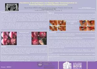
Laterality of Motor Control and Epilepsy: A Technical/Statistical Analysis of Surgical Failures, Based on the Directionality of Signals Traveling Between Hemispheres Iraj Derakhshan M.D., Charleston, W.V. Table 1. Unreliability Index (Incidences of Bilateralism, Variability) Table 2. Epilepsy Surgery Series, Side of the Operation REFERENCES 1. Derakhshan I. Laterality of motor control revisited: directionality of callosal traffic and its rehabilitation implications. Top Stroke Rehabil. 2005; 12:76-82. 2. Derakhshan I. Anatomy of handedness and the laterality of seizure onset: Surgical implications of new understandings in motor control. Neurol Res. 2005; In Press 3. Kobayashi M, Hutchinson S, Schlaug G, Pascual-Leone A. Ipsilateral motor cortex activation on functional magnetic resonance imaging during unilateral hand movements is related to interhemispheric interactions. Neuroimage. 2003; 20:2259-2270. (Fig. 4, page 2268) 4. Wada M, Yamamoto S, Kitazawa S. Effects of handedness on tactile temporal order judgment. Neuropsychologia. 2004; 42:1887-1895. 5. Duane DC, Ng YT, Rekate HL, Chung S, Bodensteiner JB, Kerrigan JF. Treatment of refractory status epilepticus with hemispherectomy.Epilepsia. 2004; 45:1001-1004. 6. Ven AA, Van Hees JG, Stappaerts KH. Effect of size and pressure of surface recording electrodes on amplitude of sensory nerve action potentials. Muscle Nerve. 2004; 30:234-238. 7. Kobayashi K, Maniwa S, Ogino T, Yoshinaga H, Ohtsuka Y, Oka E. Myoclonic seizures combined with partial seizures and probable pathophysiology of secondary bilateral synchrony. Clin Neurophysiol. 2000; 111:1813-1816. (Fig.2) 8. Blume WT. Hemispheric epilepsy. Brain. 1998; 121:1937-1949. (Table 2) 9. Kobayashi K, Nishibayashi N, Ohtsuka Y, Oka E, Ohtahara S. Epilepsy with electrical status epilepticus during slow sleep and secondarybilateral synchrony. Epilepsia. 1994; 35:1097-1103. 10. Smith MC. The utility of magnetoencephalography in the evaluation of secondary bilateral synchrony: a case report. Epilepsia. 2004; 45 Suppl 4:57-60. 11.Bingley T. Mental symptoms in temporal lobe epilepsy and temporal lobe gliomas with special reference to laterality of lesion and the relationship between handedness and brainedness; a study of 90 cases of temporal lobe epilepsy and 253 cases of temporal lobe glioma. Acta Psychiatr Neurol Scand. 1958; 33(Suppl 120):1-151. 12. Cibula JE, Gilmore RL. Secondary epileptogenesis in humans. J Clin Neurophysiol. 1997; 14:111-127. 13. Derakhshan I. Crossed nonaphasia in a dextral with left hemispheric lesions: handedness technically defined. Stroke. 2002; 33:1749-1750. 14. Crone NE, Miglioretti DL, Gordon B, Sieracki JM, Wilson MT, Uematsu S, Lesser RP. Functional mapping of human sensorimotor cortex with electrocorticographic spectral analysis. I. Alpha and beta event-related desynchronization. Brain. 1998; 121:2271-2299. 15. Mazzini L, Schieppati M. Activation of the neck muscles from the ipsi- or contralateral hemisphere during voluntary head movements in humans. A reaction-time study. Electroencephalogr Clin Neurophysiol. 1992; 85:183-189. 16. Bladin PF. Intra-carotid treatment of status epilepticus. Epilepsia. 1963; 54:151-166. 17. Park YD, Hoffman JM, Radtke RA, DeLong GR. Focal cerebral metabolic abnormality in a patient with continuous spike waves during slow-wave sleep. J Child Neurol. 1994; 9:139-143. 18. Derakhshan I. Treatment of refractory status epilepticus with hemispherectomy: A note of caution. Epilepsia. In Press 19. Gloor P, Rasmussen T, Altuzarra A, Garretson H. Role of the intracarotid amobarbital-pentylenetetrazol EEG test in thediagnosis and surgical treatment of patients with complex seizure problems. Epilepsia. 1976; 17:15-31. (CASE J. DU, Fig. 3) 20. Rovit RL, Gloor P, Rasmussen T. Intracarotid amobarbital in epileptic patients. A new diagnostic tool in clinical electroencephalography. Arch Neurol. 1961 5:606-626. (Cases 1A, a left- hander with right dominant cortex; Cases B1, B2 with “independent” foci, minor hemisphere injections) 21. Parent A. Auguste Forel on ants and neurology. Can J Neurol Sci. 2003; 30:284-291. 22. Hunter KE, Blaxton TA, Bookheimer SY, Figlozzi C, Gaillard WD, Grandin C, Anyanwu A, Theodore WH. (15) O water positron emission tomography in language localization: a study comparing positron emission tomography visual and computerized region of interest analysis with the Wada test. Ann Neurol. 1999; 45:662-665. (Patient 1) 23. Springer JA, Binder JR, Hammeke TA, Swanson SJ, Frost JA, Bellgowan PS, Brewer CC, Perry HM, Morris GL, Mueller WM. Language dominance in neurologically normal and epilepsy subjects: a functional MRI study.Brain. 1999; 122:2033-2046. (Table 4) 24. McMackin D, Jones-Gotman M, Dubeau F, Gotman J, Lukban A, Dean G, Evans A, Lisbona R. Regional cerebral blood flow and language dominance: SPECT during intracarotid amobarbital testing. Neurology. 1998; 50:943-50. 25. Rasmussen T, Milner B. The role of early left-brain injury in determining lateralization of cerebral speech functions. Ann N Y Acad Sci. 1977 30; 299:355-369. 26. Altenmuller E. Cortical DC-potentials as electrophysiological correlates of hemispheric dominance of higher cognitive functions.Int J Neurosci. 1989; 47:1-14. 27. Labar D, Dilone L, Solomon G, Harden C. Epileptogenesis: left or right hemisphere dominance? Preliminary findings in a hospital-based population. Seizure. 2001; 10:570-572. 28. Scott DF. Left and right cerebral hemisphere differences in the occurrence of epilepsy. Br J Med Psychol. 1985; 58:189-192. 29. Derakhshan I. Electroencephalogram and laterality of movement control: a clinical analysis. J Child Neurol. 2003; 18:878-879. 30. Manaut E, Gomez CM, Vaquero E, Rodriguez E. Hemispheric lateralization of language in epileptic right-handed children withunihemispheric discharge. J Child Neurol. 2002; 17:505-509.31. Sirven JI, Malamut BL, O'Connor MJ, Sperling MR. Temporal lobectomy outcome in older versus younger adults. Neurology. 2000; 54:2166-2170. Page 2167 32. Arle JE, Perrine K, Devinsky O, Doyle WK. Neural network analysis of preoperative variables and outcome in epilepsy surgery. J Neurosurg. 1999; 90:998-1004. (Table 2, Discussion) 33. McIntosh AM, Kalnins RM, Mitchell LA, Fabinyi GC, Briellmann RS, Berkovic SF. Temporal lobectomy: long-term seizure outcome, late recurrence and risks for seizure recurrence. Brain. 2004; 127:2018-2030. 34. Kossoff EH, Vining EP, Pillas DJ, Pyzik PL, Avellino AM, Carson BS, Freeman JM. Hemispherectomy for intractable unihemispheric epilepsy etiology vs outcome. Neurology. 2003; 61:887-890. 35. Yoon HH, Kwon HL, Mattson RH, Spencer DD, Spencer SS. Long-term seizure outcome in patients initially seizure-free after resective epilepsy surgery. Neurology. 2003 26; 61:445-450. 36. Amar AP, Apuzzo ML, Liu CY. Vagus Nerve Stimulation Therapy after Failed Cranial Surgery for Intractable Epilepsy: Results from the Vagus Nerve Stimulation Therapy Patient Outcome Registry. Neurosurgery. 2004; 55:1086-1093. 37. Talairach J, Bancaud J, Bonis A, Szikla G, Trottier S, Vignal JP, Chauvel P, Munari C, Chodkievicz JP. Surgical therapy for frontal epilepsies. Adv Neurol. 1992; 57:707-732. (Pages 716, 718) 38. McIntosh AM, Wilson SJ, Berkovic SF. Seizure outcome after temporal lobectomy: current research practice and findings. Epilepsia. 2001; 42:1288-1307. Page 1294 39. Matsuo A, Ono T, Baba H, Ono K. Callosal role in generation of epileptiform discharges: quantitative analysis of EEGs recorded in patients undergoing corpus callosotomy. Clin Neurophysiol. 2003; 114:2165-2171. 40. Brasil-Neto JP, Araujo VP, Carneiro CR. Postexercise facilitation of motor evoked potentials elicited by ipsilateral voluntary contraction. Muscle Nerve. 1999; 22:1710-1712. 41. Kakigi R. Ipsilateral and contralateral SEP components following median nerve stimulation: effects of interfering stimuli applied to the contralateral hand. Electroencephalogr Clin Neurophysiol. 1986; 64:246-259. 42. Goto M, Okuda J, Ikejiri Y, Nishikawa T, Hirose M, Tanabe H, Nii Y, Nakatani S, Shiraishi J. Somatosensory evoked potentials to median nerve stimulation after partial section of the corpus callosum.J Neurol. 1991; 238:161-167. 43. Villanueva J, Castilla JM. [Anatomo-functional considerations on the pathways generated by cortical somatosensory evoked potentials of the median nerve. Study in 6 hemispherectomized patients] Arch Neurobiol (Madr). 1988; 51:316-323.