Download
1 / 14
150 likes | 175 Views
Colony Morphology. Figure 7-4 Colony morphology descriptions. E. coli. medium sized colonies regular margin convex elevation. Klebsiella pneumoniae. Slightly gummy/wet looking colonies Circular convex entire margin.

E N D
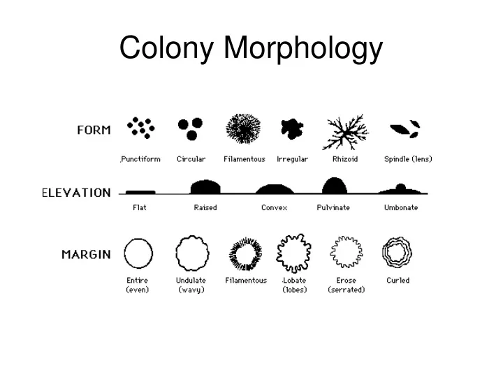
Colony Morphology Figure 7-4 Colony morphology descriptions
E. coli medium sized colonies regular margin convex elevation
Klebsiella pneumoniae Slightly gummy/wet looking colonies Circular convex entire margin
Bacillus subtilis. These gram positive, sporeforming rods produce colonies which are dry, flat, and irregular, with lobate margins.
Pseudomonas aeruginosa. This gram negative rod forms mucoid colonies with umbonate elevation. Some strains produce a diffusable green pigment and a distinctive fruity odor. P. aeruginosa is an opportunistic contaminant of burn injurys, wounds such as cuts and gunshot, and can cause bacterial pneumonia. It is often nosocomial pathogen, easily transmitted by hands and invasive medical procedures.
Salmonella choleraesuis serovar typhimurium. This gram negative rod is a component of the gastrointestinal tract of birds and reptiles and is an agent of gastroenteritis in humans. It forms shiny, convex colonies with entire margins.
Escherichia coli. This gram negative rod (coccobacillus) forms shiny, mucoid colonies which have entire margins and are slightly raised. Older colonies often have a darker center.
Rhodospirillum rubrum. Pinpoint circular colonies which are convex with entire margins. This gram negative spirillum produces a non-diffusable red pigment.
Micrococcus luteus. Circular, pinhead colonies which are convex with entire margins. This gram positive coccus produces a bright yellow, non-diffusable pigment
Staphylococcus aureus. Circular, pinhead colonies which are convex with entire margins. This gram positive coccus often produces colonies which have a golden-brown color.
Staphylococcus epidermidis. Circular, pinhead colonies which are convex with entire margins. The colonies of this gram-positive coccus appear either the color of the agar, or whitish.
Bacillus subtilis Colonies large margin is undulate or lobate dry with circular form flat elevation
Staphylococcus aureus. Circular Pinhead Convex Entire margin
Serratia marcescens Mucoid Entire margin Umbonate elevation Red and white colonies