Download
1 / 21
220 likes | 384 Views
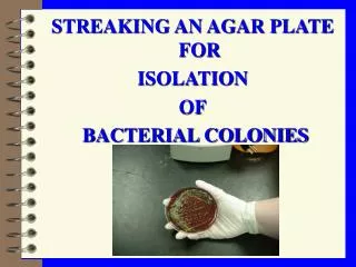
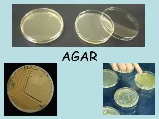
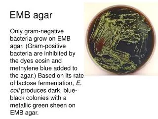
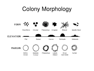
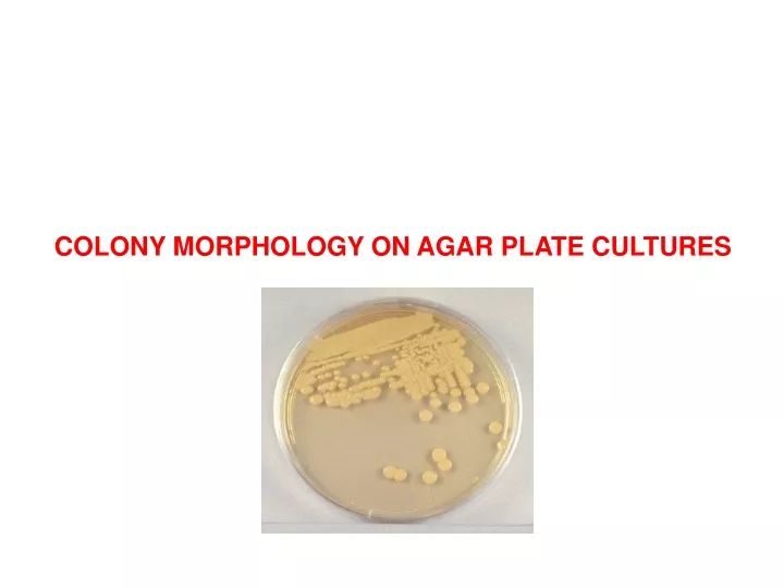
COLONY MORPHOLOGY ON AGAR PLATE CULTURES. Form of Colony. Elevation of Colony. Margin of Colony. Surface of Colony. There are three common shapes of bacteria. 1-Coccus. having one of the following arrangements. Diplococcus: a pair of cocci. Streptococcus: a chain of cocci.

E N D
There are three common shapes of bacteria 1-Coccus having one of the following arrangements Diplococcus: a pair of cocci Streptococcus: a chain of cocci Tetrad: a square of 4 cocci Sarcina: a cube of 8 cocci Staphylococcus: cocci in irregular, often grape-like clusters
Bacillus (rod)2- Bacillus: a single bacillus Streptobacillus: bacilli in chains Coccobacillus: oval and similar to a coccus
Spiral3- Vibrio: an incomplete spiral or comma-shaped Spirillum: a thick, rigid spiral Spirochete: a thin, flexible spiral
Simple Stai Simple Staining and Bacterial Cell Morphology
Preparing a smear for staining.(The following procedure is used for all of our staining) • 1.Flame (sterilize) your inoculating loop/needle before and after use.
2. Prepare the smear • If you have a solid culture (agar colony), • place a small drop of water on a clean slide. • Drag the sterile inoculating needle tip • through the edge of colony. b. Gently spread the mixture into a circle. A loop of liquid culture can be placed directly on the slide and spread out.
4.Heat-Fix the smear. Smears are heat-fixed by quickly passing the slide through a flame two or three times. This causes the microbes to stick to the slide and not get washed off during the staining process.
5.Stain the smear. Place the slide on a rack over the sink. Flood the smear with stain and let it for 60-90 seconds. Rinse gently and blot dry.
6. Observe the slide under low and high-dry lenses to locate, center, and focus the image. Then, place a drop of oil directly on the stained smear .Turn the oil lens into position and fine focus to observe the cells.
Name the bacterial morphologies (shapes and arrangements) seen here. 3 1 6 2 5 4 Answers: 1. Spirillum 2. Coccus 3. Bacillus 4. Diplobacillus 5. Streptobacillus 6. Diplococcus
Aspergillus sp. Penicillium sp.