Download
1 / 101
1.06k likes | 1.75k Views
Chapter 8: Recombinant DNA Technology and Molecular Cloning.

E N D
Chapter 8: Recombinant DNA Technology and Molecular Cloning
Sometimes a good idea comes to you when you are not looking for it. Through an improbable combination of coincidences, naiveté and lucky mistakes, such a revelation came to me one Friday night in April, 1983, as I gripped the steering wheel of my car and snaked along a moonlit mountain road into northern California’s redwood country. That was how I stumbled across a process that could make unlimited numbers of copies of genes, a process now known as the polymerase chain reaction (PCR) Kary B. Mullis, Scientific American (1990), 262:36
The cornerstone of most molecular biology technologies is the gene. • To facilitate the study of a genes: • Clone the gene by inserting it into another DNA molecule that serves as a vehicle or vector that can be replicated in living cells.
When two DNAs (the insert and vector) of different origin are combined, the result is a recombinant DNA molecule. • The recombinant DNA is placed in a host cell, amplified, and purified for further analysis.
Recombinant DNA technology arose through the efforts of several research groups working primarily on bacteriophage lambda ().
Insights from bacteriophage lambda () cohesivesites • In 1962, Allan Campbell noted that the linear genome of bacteriophage forms a circle upon entering the host bacterial cell by joining complementary single-stranded DNA cohesive (cos) sites. • The idea of joining DNA segments by “cohesive sites” became a guiding principle in the development of recombinant DNA technology.
Insights from bacterial modification and restriction systems Salvador Luria and other phage workers made the following observations: • Phages grown in one bacterial host fail to grow in a different “restrictive” bacterial host. • The phage DNA is degraded in the “restrictive” host.
Rare progeny phages become “modified” in some way so that they grow normally in the new host. • The modification was reversible. • 1962: The molecular basis of restriction and modification was defined by Werner Arber and coworkers.
Restriction system Restriction endonucleases • First restriction endonuclease characterized in E. coli K-12 by Matt Meselson and Bob Yuan. • “Restrict” or prevent viral infection by degrading the invading nucleic acid.
Modification system • Methylase activity: Addition of methyl groups to protect those sites in DNA sensitive to attack by a restriction endonuclease. • Typically adenine methylation (6-methyl adenine). • Methylation pattern is maintained during DNA replication.
The first cloning experiments • One of the first recombinant DNA molecules was a hybrid of phage and the SV40 mammalian DNA virus genome. • 1974: first eukaryotic gene was cloned. • Amplified ribosomal RNA (rRNA) genes from Xenopus laevis oocytes were cloned into a bacterial plasmid. • The cloned frog genes were actively transcribed into rRNA in E. coli.
I was tempted then to put together a book called the Whole Risk Catalogue. It would contain risks for old people and young people and so on. It would be a very popular book in our semi-paranoid society. Under “D” I would put dynamite, dogs, doctors, dieldrin [an insecticide] and DNA. I must confess to being more frightened of dogs. But everyone has their own things to worry about. James Watson, Genetics and Society (1993)
Fear of recombinant DNA molecules • 1975: Recommendations from a landmark meeting of molecular biologists formed the basis for official guidelines developed by the National Institutes of Health (NIH). • Activities involving the handling of recombinant DNA and organisms must be conducted in accordance with the NIH guidelines. • Four levels of risk are recognized, from minimal to high.
Two main categories of enzymes are important tools in the preparation of recombinant DNA • DNA ligases: join two pieces of DNA by forming phosphodiester bonds. • Restriction endonucleases: recognize a specific, rather short, nucleotide sequence on a double-stranded DNA molecule, called a restriction site, and cleave the DNA at this site or elsewhere.
Major classes of restriction endonucleases • Type II restriction endonucleases are widely used by molecular biologists. • >240 available commercially. • 6 bp cutters are the most commonly used.
Restriction endonucleases are named for the organism in which they were discovered: • HindIII from Haemophilus influenza (strain d) • SmaI from Serratia marcescens • EcoRI from Escherichia coli (strain R) • BamHI from Bacillus amyloliquefaciens (strain H)
Recognition sequences for type II restriction endonucleases • Orthodox type II restriction endonucleases function as homodimers. • Recognition sequences are typically palindromes. • Some enzymes generate “sticky ends.” • Some enzymes generate “blunt ends.”
Restriction endonucleases exhibit a great degree of sequence specificity. • A single base pair change in the recognition site eliminates enzymatic activity.
The steps involved in restriction endonuclease DNA binding and cleavage • The first contact is nonspecific binding: • Interaction with the DNA sugar-phosphate backbone only. • Catalytic center kept at a safe distance.
Random walk: • “Sliding” over short distances of <30-50 bp to target restriction site. • “Hopping” or “jumping” over longer distances. • Specific binding at restriction site: • Large conformational change of the enzyme and DNA (coupling). • Activation of catalytic center.
EcoRI: kinking and cutting DNA • Common structural core of four conserved -strands and one -helix. • Large conformational change in EcoRI and the DNA upon specific binding. • A central kink in the DNA brings the critical phosphodiester bond between G and A deeper into the active site and unwinds the DNA.
In the presence of Mg2+, EcoRI cleaves the DNA on both strands at the same time to give free 5′-phosphate and 3′-OH ends. • The exact mechanism by which cleavage occurs has not yet been proven experimentally.
DNA ligase joins linear pieces of DNA • The DNA ligase most widely used in the lab is from bacteriophage T4. • T4 DNA ligase catalyzes formation of a phosphodiester bond between the 5′-phosphate of a nucleotide on one fragment of DNA and the 3′-hydroxyl of another.
T4 DNA ligase will ligate fragments with sticky ends or blunt ends, but for blunt ends the reaction is less efficient. • To increase the efficiency of ligation, researchers often use the enzyme terminal deoxynucleotidyl transferase to modify the blunt ends.
Basic molecular cloning procedure • DNA fragments to be cloned are generated using restriction endonucleases. • Fragments are ligated to other DNA molecules that serve as vectors. • Recombinant DNA molecules are transferred to a host cell. • Cloned recombinant DNA is recovered from the host cell for analysis.
Choice of vector is dependent on insert size and application Cloning vectors are carrier DNA molecules with four important features: • Replicate independently. • Contain a number of restriction endonuclease cleavage sites that are present only once. • Carry a selectable marker. • Relatively easy to recover from host cell.
The greatest variety of cloning vectors has been developed for use in E. coli. • The first practical skill generally required by a molecular biologist is the ability to grow pure cultures of bacteria.
Classic cloning vectors: • Plasmids • Phages • Cosmids New generation vectors: • Bacterial artificial chromosomes (BACs) • Yeast artificial chromosomes (YACs) • Mammalian artificial chromosomes (MACs)
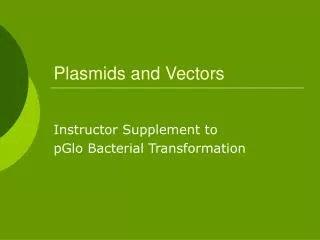
Plasmid DNA as a vector • Plasmids are named with a system of uppercase letters and numbers, where the lowercase “p” stands for “plasmid.” • Low copy number plasmids: replicate to yield only one or two copies in each bacterial cell. • High copy number plasmids: replicate to yield >500 copies per bacterial cell.
Plasmid vectors are modified from naturally occurring plasmids • Contain a specific antibiotic resistance gene. • Contain a multiple cloning site.
Five major steps for molecular cloning using a plasmid vector • Construction of a recombinant DNA molecule. • Transfer of ligation reaction products to host bacteria. • Multiplication of plasmid DNA molecules. • Division of host cells and selection of recombinant clones, e.g. by blue-white screening. • Amplification and purification of recombinant plasmid DNA.
Transformation: transfer of recombinant plasmid DNA to a bacterial host • Bacterial cells are incubated in a concentrated calcium salt solution to make their membranes leaky. • The permeable “competent” cells are mixed with DNA to allow DNA entry. • Alternatively, a process called electroporation drives DNA into cells by a strong electric current.
Why isn’t the introduced foreign plasmid DNA degraded by a bacterial restriction-modification system?
Recombinant selection • Antibiotic resistance selects for transformed bacterial cells. • Numerous cell divisions of a single transformed bacteria result in a clone of cells visible as a bacterial colony on an agar plate. • Successfully transformed bacteria will carry either recombinant or nonrecombinant plasmid DNA.
Blue-white screening • In the case of the vector pUC18, blue-white screening is used to distinguish recombinant from nonrecombinant transformants. • Also known as “lac selection” or - complementation
-galactosidase activity can be used as an indicator of the presence of foreign DNA • If the lacZ 5′ region of pUC18 is not interrupted by inserted foreign DNA, the amino-terminal portion of -galactosidase is synthesized. • The mutant E. coli host encodes only the carboxyl end of -galactosidase.
The N-terminal and C-terminal fragments come together to form a functional enzyme. • -galactosidase activity can be measured using a colorless chromogenic substrate called X-gal. • Cleavage of X-gal produces a blue-colored product, visualized as a blue colony on an agar plate. • If a foreign insert has disrupted the lacZ 5′ coding sequence, X-gal is not cleaved and the bacterial colonies remain white.
Amplification and purification of recombinant plasmid DNA • Further screening to confirm the presence and orientation of the insert. • Amplify positive (white) colony containing recombinant plasmid DNA in liquid culture. • Purify plasmid DNA from crude cell lysates, e.g. by chromatography and ethanol precipitation.
Liquid chromatography • Molecules dissolved in a solution will interact (bind and dissociate) with a solid surface. • When the solution is allowed to flow across the surface, molecules that interact weakly with the solid surface will spend less time bound to the surface and will move more rapidly. • Commonly used to separate mixtures of nucleic acids and proteins.
Three main techniques • Gel filtration chromatography: separation by differences in mass. • Ion-exchange chromatography: separation by differences in charge. • Affinity chromatography: separation by differences in binding affinity.
Bacteriophage lambda () as a vector • Phage vectors are particularly useful for preparing genomic libraries. • The recombinant viral particle infects bacterial host cells in a process called transduction. • Progeny viral particles appears as a clear spot of lysed bacteria or “plaque” on a lawn of bacteria.
Artificial chromosome vectors • Bacterial artificial chromosomes (BACs) and yeast artificial chromosomes (YACs) are important tools for mapping and analysis of complex eukaryotic genomes. • 1997: first prototype mammalian artificial chromosome (MAC)
Yeast artificial chromosome (YAC) vectors YAC vectors are designed to act like chromosomes in host yeast cells • Origin of replication (Autonomously replicating sequence, ARS) • Centromere • Telomere
YAC vectors contain selectable markers • URA3: encodes an enzyme required for uracil biosynthesis. • TRP1: encodes an enzyme required for tryptophan biosynthesis. • SUP4: tRNA that suppresses the Ade2-1 UAA mutation.
Red-white selection Host yeast strain: ura3/trp1/Ade2-1 mutant • When foreign DNA is inserted in the multiple cloning site, SUP4 expression is interrupted. • The Ade2-1 mutation is no longer suppressed. • ADE1 and ADE2 encode enzymes involved in adenine biosynthesis.
Ade2-1 mutant cells produce a red pigment from polymerization of an intermediate compound. • In the absence of foreign DNA, SUP4 is expressed. • The Ade2-1 mutation is suppressed. • Ade2-1 mutant cells expressing SUP4 are white (the color of wild-type yeast cells).