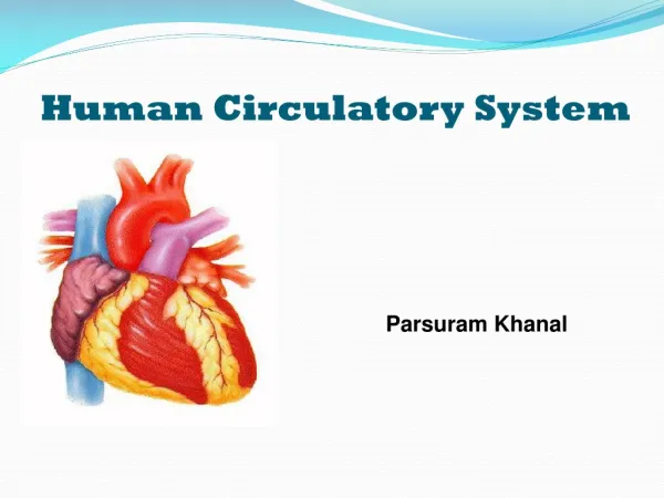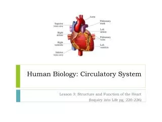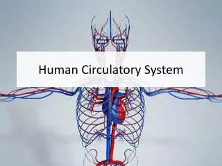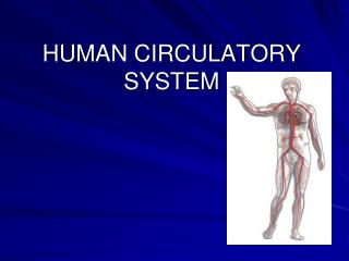
Human Circulatory System
Explore the human circulatory system, its functions, components like blood and plasma, blood types, heart structure and function, coronary circulation, and systemic circuits in maintaining the body's balance. Learn about blood vessels and the heart cycle, understanding the importance of maintaining a healthy circulatory system for overall well-being.

Human Circulatory System
E N D
Presentation Transcript
Human Circulatory System • Interacts with other body systems to maintain homeostatsis • Delivers nutrients, oxygen to cells • Carries away waste products and CO2 • Transports chemical messengers through out body • Helps maintain a constant body temperature • Plays role in blood pressure control
Structure of Circulatory System • Is made up of three basic parts: • Pump (heart) • Circulating fluid (blood) • Blood vessels to carry circulating fluid
Blood • Average adult has 4 to 6 L of blood • Is made up of four basic components • Plasma • Erythrocytes (red blood cells) • Leucocytes (white blood cells) • Platelets
Plasma • Liquid component of blood • Accounts for 55% of blood volume • Is 90% water • Contains dissolved salts, proteins, hormones, nutrients, waste products and gases
Erythrocytes • Are the most abundant cells in blood • Are manufactured in the bone marrow and stored in the spleen • Primary function is to carry oxygen • Contain no nuclei or mitochondria but do contain hemoglobin molecules • Iron is a key component of RBC giving blood its bright red colour when exposed to oxygen • RBC have a biconcave shape making them flexible and allows them to move through vessels of different shapes and sizes
Platelets • Are important agents in the blood for clotting • Are cell fragments broken from special cells in bone marrow and have no nucleus • Release chemicals (clotting factors) when they encounter damaged blood vessels • Strand-like molecules called fibrin form a mesh or clot
Leucocytes • Are out numbered by RBC’s 700 to 1 • Are responsible for helping defend the body from disease and infection • Amoeboid-shaped cells destroy and consume invading bacteria and damaged cells • There are two types both have a nucleus and granulocytes have granules in the cytoplasm while agranulocytes do not • Both are manufactured in the bone marrow
Blood Types • Proteins on the surface of the RBC determine blood type (agglutinogens A and B) • Type A has agglutinogen A • Type B has agglutinogen B • Type AB has agglutinogens A and B • Type O has no proteins on surface
Each blood type is also associated with specific proteins in the plasma called agglutinins • Type A has agglutinin B • Type B has agglutinin A • Type AB has neither • Type O has agglutinin A and B • When the same agglutinin and agglutinogen combine the blood thickens and will not flow which is lethal
Pulmonary and Systemic Circuits • The pulmonary circuit is a low-pressure system • Deoxygenated blood from the right side of the heart travels to the lungs where gas exchange occurs and then oxygenated blood returns to the left side of the heart • The systemic circuit is a high pressure system • Oxygenated blood from the left side of the heart and travels to all parts of the body
Coronary Circulation • The heart has a special circulation which provides a fresh supply of oxygen to the muscles of the heart • Blockage of a coronary artery can result in lack of oxygen to heart muscle tissue resulting in tissue death • This is a heart attack, a heart attack can also cause irregular heartbeat and prevent the heart from filling and contracting normally
Heart Cycle • Heartbeat is result of coordinated contraction of heart muscle • A region of muscle in right atrium called sinoatrial node or pacemaker maintains the heart’s intrinsic pumping rhythm • Contractions in the SA node travel to the atrioventricular node and then travel along the Purkinje fibres in the septum toward the ventricles
Heart Muscle • Cardiac muscle is composed of striated like other muscle tissue but has a unique branching pattern • Cardiac muscle is myogenic muscle because it has the ability to contract without being stimulated by external nerves which is why it can continue to beat for a short time after being removed from the body
Heart Sounds • The typical lubb-dubb heart sounds are caused by the closing of the heart valves • The heart works in a continuous cycle of relaxation and contraction • During distole the heart is relaxed and blood flows into all four chambers • Distole ends with the contraction of the atria • During this stage blood pressure is reduced and is called diastolic pressure
Systole begins with the contraction of the ventricles which forcefully expels blood from the heart • Blood passes through the semi-lunar valves into the pulmonary artery and the aorta • The tricuspid and bicuspid valves are closed to prevent blood from flowing back into the atria • Blood pressure is increased and is called the systolic pressure
Blood Vessels • Transport the blood from one part of the body to another • Blood is carried away from the heart in arteries which branch out into arterioles and then into capillaries • Arteries are thick walled into order to withstand the pressure of the blood within them • The expansion and contraction of the arteries can be felt as your pulse
Capillaries are the narrowest of all blood vessels • RBC must squeeze through in single file and so they slow down allowing them to pick up CO2 and release O2 • Every tissue of the body is within approx. 0.1 mm of a capillary • Capillaries are the bridge between the arterial and venous systems
Capillaries merge to form venules which in turn merge to form veins • Veins are reservoirs that hold about half of the total blood volume • Veins have thinner walls, larger diameters and less muscle than arteries • Veins transport blood back to the heart and must work against gravity • Blood pressure is low in the veins and so one way valves and the contraction of skeletal muscle move blood through veins