Download
1 / 17
170 likes | 206 Views
Explore the anatomy and functions of the human circulatory system, including the role of blood, pumps, vessels, and specialized organs in nutrient exchange and temperature regulation. Learn about the heart's contractions, nervous impulses, cardiac cycle, and auxiliary mechanisms that regulate cardiac output. Discover how muscular work influences cardiac output and the intricacies of the respiratory system, gas exchange, and the mechanism of breathing.

E N D
Anatomy of human circulatory system Lecture note IE 665 Applied Industrial Ergonomics
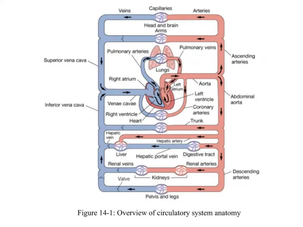
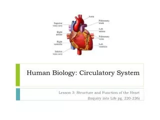
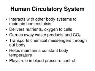
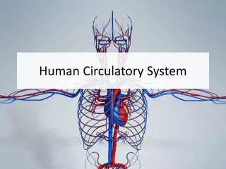
Anatomy of Human Circulatory System Function: Tomove nutrients, gases and wastes to and from cells throughout the body, and to stabilize body temperature, pH such that cells can carry out their functions. • The Main Features • Blood, to transport, nutrients, wastes, oxygen, carbon dioxide and hormones. • Two pumps (in a single heart) • one to pump deoxygenated blood to the lungs; • the other to pump oxygenated blood to all the other organs and tissues of the body. • A system of blood vessels to distribute blood throughout the body • Specialized organs for exchange of materials between the blood and the external environment; for example lungs, lever, intestine, and kidneys IE 665
The heart and its working Working of the heart valves IE 665
Nervous impulses and contraction The heart's contractions (both rate and strength) are controlled by nervous impulses generated from specialized auto-rhythmic electrical cells (nodes) located in the heart’s wall. The nerve impulses originate in the sinoatrial (SA) node, which is also known as the pacemaker, because it sets the heart's contraction rate. The impulses that it creates cause both atria to contract. The impulse then spreads to the atrioventricular (AV) node. The AV node delays the impulse for about 0.07 seconds to allow the atria to completely contract and empty. The impulse then travels from the AV node to the atrioventricular bundle, to Purkinje fibers to the cells in the ventricles, causing them to contract. This pushes blood out of the heart. After each contraction , the cardiac muscles relax passively, when heart chambers are re-filled with blood from the systemic and pulmonary circulation. Rhythmical contraction and relaxation of heart muscle are called systole and diastole. IE 665
Auxiliary mechanisms that regulate the cardiac output The rate and strength of heart’s beating can be modified by two auxiliary control centers located in the medulla oblongata of the brain. One sends nerve impulses down sympathetic (accelerans) nerves. It increases the rate and strength of the heart contraction and thus increases the cardiac output. Its activation usually arises from some stress such as fear or violent exertion. The heartbeat may increase to 180 beats per minute. The strength of contraction increases as well so the amount of blood pumped may increase to as much as 25-30 liters/minute. The other sends nerve impulses down a pair of parasympathetic (vagus) nerves. They, too, run from the medulla oblongata to the heart. Their activity slows the heartbeat. Pressure receptors in the aorta and carotid arteries send impulses to the medulla which relays these — by way of the vagus nerves — to the heart. Heartbeat and blood pressure diminish. IE 665
How muscular work influences cardiac output (1) As cellular respiration increases, so does the carbon dioxide level in the blood. This stimulates receptors in the carotid arteries and aorta, and these transmit impulses to the medulla for relay — by the sympathetic nerve — to the heart. (2) As muscular activity increases, the muscle pump drives more blood back to the right atrium. The atrium becomes distended with blood, thus stimulating stretch receptors in its wall. These, too, send impulses to the medulla for relay to the heart. IE 665
Respiratory system When a breath is taken, air passes in through the nasal passages, into the pharynx, through the larynx, down the trachea, into the right and left bronchi, which branches and rebranches into bronchioles, each of which terminates in a cluster of alveoli Inside the lung. It is here that gas exchange occurs. There are about 300 million alveoli, which together provide a very large surface area (equivalent to a tennis courts and 80 times our skin area); plenty surface area for gas exchange. A rubber cast of human lungs (courtesy of the Anatomical Institute, Bern) IE 665
The act of breathing • During inspiration (inhaling), • The external intercostal muscles contract, lifting the ribs up and out. • The diaphragm contracts, drawing it down . Chest cavity is increased and negative pressure in alveoli draws in air. • During expiration (exhaling), these processes are reversed and the natural elasticity of the lungs returns them to their normal volume. • At rest, we breath 15-18 times a minute exchanging about 500 ml of air. • In more vigorous expiration, • The internal intercostal muscles draw the ribs down and inward • The wall of the abdomen contracts pushing the stomach and liver upward. • Under these conditions, an average adult male can flush his lungs with about 4 liters of air at each breath. This is called the vital capacity. Even with maximum expiration, about 1200 ml of residual air remain. IE 665
Alveolar capillary bed and gas exchange The pulmonary artery, carrying carbon dioxide rich blood from the left ventricle subdivides many times and forms a rich alveolar capillary bed around alveoli. The tissue separation between alveoli and capillaries are extremely thin. Oxygen and carbon dioxide are exchanged between blood and the air by diffusion and difference in partial pressure. Blood takes up oxygen and discharges carbon dioxide. This blood then flows out of the alveolar capillaries, through venuoles, and back to the heart via the pulmonary veins. Hemoglobin pigment in the red blood cell is the main constituent that facilitates blood to carry oxygen. Electron micrograph of two alveoli (Air) and an adjacent capillary from the lung of a laboratory mouse. Note the thinness of the epithelial cells (EP) that line the alveoli and capillary. At the closest point, the surface of the red blood cell is only 0.7 µm away from the air in the alveolus. IE 665
Control of Breathing • The rate of cellular respiration (and hence oxygen consumption and carbon dioxide production) varies with level of activity. Vigorous exercise can increase by 20-25 times the demand of the tissues for oxygen. This is met by increasing the rate and depth of breathing. • It is a rising concentration of carbon dioxide — not a declining concentration of oxygen — that plays the major role in regulating the ventilation of the lungs. The concentration of CO2 is monitored by cells in the medulla oblongata. If the level rises, the medulla responds by increasing the activity of the motor nerves that control the intercostal muscles and diaphragm. • However, the carotid body in the carotid arteries does have receptors that respond to a drop in oxygen. Their activation is important in situations (e.g., at high altitude in the unpressurized cabin of an aircraft) where oxygen supply is inadequate but there has been no increase in the production of CO2. • The smooth muscle in the walls of the bronchioles is very sensitive to the concentration of carbon dioxide. A rising level of CO2 causes the bronchioles to dilate. This lowers the resistance in the airways and thus increases the flow of air in and out. IE 665
Systemic circulation and blood pressure • Blood moves through the systemic arteries, arterioles, and capillaries because of the force/pressure created by the contraction of the ventricles. The surge of blood that occurs at each contraction is transmitted through the elastic walls of the entire arterial system where it can be detected as the pulse. Even during the brief interval when the heart is relaxed — called diastole — there is still pressure in the arteries. Blood pressure is expressed as two numbers, e.g., 120/80. • The first is the pressure during systole, the pressure equivalent to that produced by a column of mercury 120 mm high. The second number is the pressure at diastole. • Blood pressure in the veins • When blood leaves the capillaries and enters the venules and veins, little pressure remains to force it along. Blood in the veins below the heart is helped back up to the heart by the muscle pump. This is simply the squeezing effect of contracting muscles on the veins running through them. One-way flow to the heart is achieved by valves within the veins. • With muscular work, strength and rate of heart contraction increases with concurrent increase of blood pressure. • Although blood pressure can vary greatly in an individual, continual high pressure — especially diastolic pressure — may be the symptom or cause of a variety of ailments. The medical term for high blood pressure is hypertension. IE 665
Control of the Capillary Beds • An adult human has been estimated to have some 60,000 miles of capillaries with a total surface area of some 800–1000 m2 (an area greater than three tennis courts). The total volume of this system is roughly 5 liters, the same as the total volume of blood. However, if the heart and major vessels are to be kept filled, all the capillaries cannot be filled at once. So a continual redirection of blood from organ to organ takes place in response to the changing needs of the body. During vigorous exercise, for example, capillary beds in the skeletal muscles open at the expense of those in the viscera. The reverse occurs after a heavy meal. Heat stress significantly reduced Capillary Blood Flow to inner body organs, by shunting more blood to skin capillaries for dissipation of heat. • The walls of arterioles are encased in smooth muscle. Constriction of arterioles decreases blood flow into the capillary beds they supply while dilation has the opposite effect. In time of danger or other stress, for example, the arterioles supplying the skeletal muscles will be dilated while the bore of those supplying the digestive organs will decrease. • This redistribution of blood is controlled by- • Autonomous (sympathetic and parasympathetic) nervous system causing dilation or constriction of the arteriols’ diameter by acting on the smooth muscles of the arteriols walls. • Local control. Presence of metabolites and other chemicals concentration to dilate the capillary diameter. IE 665
Redistribution of blood during vigorous exercise The table shows the distribution of blood in the human body at rest and during vigorous exercise. Note the increase in blood supply to the working organs (skeletal muscles and heart). The increased blood supply to the skin aids in the dissipation of the heat produced by the muscles. Note also that the blood supply to the brain remains constant. The total blood flow during exercise increases because of a more rapid heartbeat and also a greater volume of blood pumped at each beat. IE 665
Cellular respiration • When blood enters the arteriole end of a capillary, it is still under pressure (about 35 torr) produced by the contraction of the ventricle. As a result of this pressure, a substantial amount of water and some plasma proteins filter through the walls of the capillaries into the tissue space. • Interstitial fluid bathes the cells in the tissue space and substances in it can enter the cells by diffusion and active transport. Substances, like oxygen, carbon dioxide, can diffuse in and out of cells and into the interstitial fluid. • Near the venous end of a capillary, the blood pressure is greatly reduced (to about 15 torr). Here another force comes into play. Although the composition of interstitial fluid is similar to that of blood plasma, it contains a smaller concentration of proteins than plasma and thus a somewhat greater concentration of water. This difference sets up an osmotic pressure (~ 25 torr), which is greater than the blood pressure at the venous end of the capillary. Consequently, the fluid reenters the capillary here. IE 665