Download
1 / 58
590 likes | 731 Views
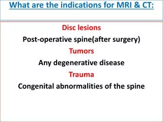
What are the indications for MRI & CT:. Disc lesions Post-operative spine(after surgery) Tumors Any degenerative disease Trauma Congenital abnormalities of the spine. Preparations for CT & MRI: 1-Fasting for 4-6 hours 2-Contrast material Urographin ,telebrix 1-2 mg/kg

E N D
What are the indications for MRI & CT: Disc lesions Post-operative spine(after surgery) Tumors Any degenerative disease Trauma Congenital abnormalities of the spine
Preparations for CT & MRI:1-Fasting for 4-6 hours2-Contrast material Urographin ,telebrix 1-2 mg/kg • Administration of contrast: Yes: In post-operative lumbar spine, inflammatory disorders& neoplastic lesions. No:disc lesions, trauma. Anesthesia Used with children & uncooperative patients.
Patient position: Is usually supine, sometimes side lying, & rarely prone. How to see: Scanogram: primitive picture to detect the site of lumbar spine
Computed tomography (CT): . Usual scanning. . Axial slides 2-4 mm. 2 mm in cervical spine / 4 mm in lumbar spine Has bone & soft tissue window. CT screening: . Whole segment of the spine 5 mm in cervical spine, 8 mm in lumbar spine. . Selective Scanning Every 3 mm especially in trauma, also in cervical disc lesions. N.B: If I want to see one vertebra e.g. L3 I have to take 1 vertebra above (L2 )& 1 below (L4).
CT Myelography: • Is considered as intrathecal contrast injection with L- puncture needle. • We have 2 windows • Soft tissue & bone window. What are the structures I should evaluate in CT of lumbar spine: • Lumbar spinal canal diameter normally 13 mm. • Disc lesions. • Others. • facet, sacroiliac joints & paravertebral soft tissue.
1. Lumbar spinal canal diameter: Spinal canal is bony structure, so we see it in bone window. The spinal canal must be closed (at the level of pedicles). We measure the AP diameter. Types of canal stenosis: 1-Relative: 11-12 mm & this doesn’t need operation but it needs operation if there’s disc. 2- Absolute: 8 -10 mm & it must be operated.
Disc lesions: We detect it in soft tissue window. Posterior border of the disc is more important as it has relation to the disc. The normal posterior border of the disc is CONCAVE. The abnormal is STRAIGHT OR CONVEX.
N.B: . Normally due to overload ,the disc of L5-S1 is CONVEX& the abnormal is also convex,so to judge if it’s normal or no look at the next slide if: the posterior border of the disc is convex so it is ABNORMAL. . The angle of inclination in L5-S1 is more than 30 & the device accept up till 30 only so part of the slide will contain bone & part will contain disc.
MRI – LUMBAR SPINEM NERVE ROOTS FORAMEN AXIAL VIEW
Manifestations of arthritis in any joint: (Spondylosis in spine and osteoarthritis of other joints) • Osteophytic lipping. • Narrow joint space. • Subarticular bone sclerosis • Sub cortical pseudo cystic changes. • Intra articular air.(vaccum phenomena)
We have 2 types of joints: • Neurocentral joint: Is the articulation between one vertebra above & one vertebra below which makes the shape of the body of the vertebral end plate . • Facet joint: Is the articulation between the inferior lip of the transverse process of one vertebra above with the superior lip of the transverse process of the vertebra below, it’s called Hamburger’s Sandwich. Arthritis of the neurocentral or facet joint gives the same manifestations of nerve compression due to disc lesion.
Cervical disc in CT: • We see it in soft tissue window .The disc in cervical spine is very narrow so every slide will contain both disc & bone, therefore there isn’t a slide of pure disc, So we choose the slide which contain more disc for assessment. . All posterior edges of cervical spine are normally convex. . See if there’s disc substance protruded than the bone. . Normal spinal cord picture is kidney shaped.
CT– CERVICAL SPINE C2-3 INTERVERTEBRAL DISC FORAMEN DISC FORAMEN FACET JOINT FACET JOINT SPINOUS PROCESS AXIAL SAGITTAL
CT- CERVICAL SPINE C-1 SECTION ARCH OF C-1 DENS BASE OF SKULL MASTOID AXIAL SAGITTAL
CT- LUMBAR SPINE POST MYELOGRAM DISC SPINOUS PROCESS
CT– CERVICAL SPINE C- 3 SECTION PEDICLE PEDICLE LAMINA AXIAL SAGITTAL
CT-- CERVICAL SPINE C-2 SECTION C-2 BODY DEGENERATED C6-7 C-2 SPINOUS PROCESS AXIAL SAGITTAL
CT- LUMBAR SPINE POST MYELOGRAM Axial PEDICLE PEDICLE NERVE ROOTS
CT- LUMBAR SPINE POST MYELOGRAM FORAMEN FORAMEN