Download
1 / 23
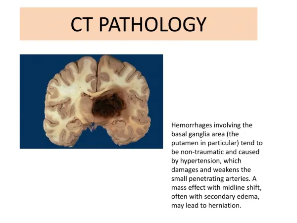
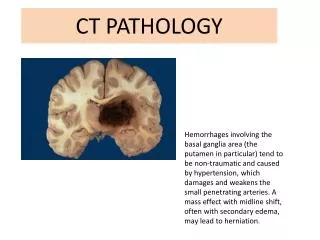
230 likes | 524 Views
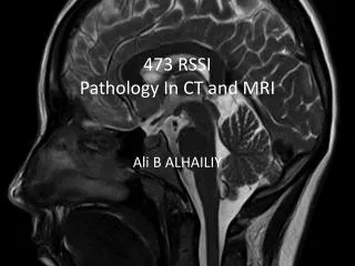
473 RSSI Pathology In CT and MRI. Ali B ALHAILIY. Course content. RSSI 473 Pathology In CT and MRI Credits 3 units (2+1) This course provides through coverage of common disease diagnosable via CT and MRI

E N D
473 RSSIPathology In CT and MRI Ali B ALHAILIY
Course content • RSSI 473 • Pathology In CT and MRI • Credits 3 units (2+1) • This course provides through coverage of common disease diagnosable via CT and MRI • Emphasis is placed on examination and demonstration of each disease or trauma process .
Focusing on following • Disease description • Disease etiology • Associated symptoms • Diagnosis • Appearance on CT and MRI
Co-ordinator’s/ Lecturer: Mr. Ali Alhailiy • Phone: 0556050106 • Office: Radiology Staff office • Email: a.alhailiy@sau.edu.sa
Learning and Teaching Activities • Lectures - didactic delivery of information to groups • Problem based learning - learning centered around a clinical situation with triggers • Online resources - content or learning activities available via the internet. • Independent and group study - work often done outside of the classroom either individually or in a group. • Assignment - written presentation that is a means of learning and assessment • Multiple choice quiz - a measurement of student knowledge and understanding performed during semester • End of semester examination - a formal measurement of student knowledge and understanding performed during the examination period of the semester.
Practical Attendance • Practical or tutorial attendance will be recorded for all MRS units of study. Students are expected • to participate in a minimum of 80% of practicals or tutorials in any unit of study. If there is any • dispute about student assessment or request for special consideration, extension or deferment, • practical attendance will be taken into account.
Required Textbooks: • It is a requirement of the unit of study that you purchase the texts listed under the required common textbooks and your discipline specific text. • 1.Sectional Anatomy For Imaging Professionals (Lorrie .l kelley) • http://www.amazon.com/Sectional-Anatomy-Imaging-Professionals-2e/dp/0323020038 • 2.Cross Sectional Anatomy CT & MRI (BhavinJhankaria) • http://www.barnesandnoble.com/w/cross-sectional-anatomy-ct-and-mri-g-bhavin-jankharia/1104348580?ean=9789350250464
kidney stone(Renal Calculi ) • A kidney stone is a solid piece of material that forms in a kidney when substances that are normally found in the urine become highly concentrated. • LocationKidney stones usually are formed inside the kidney, but they are sometimes found in the bladder or ureter. • A stone may stay in the kidney or travel down the urinary tract. Kidney stones vary in size. A small stone may pass on its own, causing little or no pain. A larger stone may get stuck along the urinary tract and can block the flow of urine, causing severe pain or bleeding.
Who gets kidney stones? Anyone can get a kidney stone, but some people are more likely to get one. Men are affected more often than women. Overweight and obese people are more likely to get a kidney stone than people of normal weight. What causes kidney stones? Kidney stones can form when substances in the urine—such as calcium, oxalate, and phosphorus—become highly concentrated. Certain foods may promote stone formation in people who are susceptible, but scientists do not believe that eating any specific food causes stones to form in people who are not susceptible. People who do not drink enough fluids may also be at higher risk, as their urine is more concentrated.
What are the types of kidney stones? Four major types of kidney stones can form: • Calcium stones are the most common type of kidney stone and occur in two major forms: calcium oxalate and calcium phosphate. Calcium oxalate stones are more common. • Calcium oxalate stone formation may be caused by high calcium and high oxalate excretion. • Calcium phosphate stones are caused by the combination of high urine calcium and alkaline urine, meaning the urine has a high pH. 2. Uric acid stones form when the urine is persistently acidic. A diet rich in purines—substances found in animal protein such as meats, fish, and shellfish—may increase uric acid in urine. If uric acid becomes concentrated in the urine, it can settle and form a stone by itself or along with calcium. 3. Struvitestones result from kidney infections. Eliminating infected stones from the urinary tract and staying infection-free can prevent more struvite stones. 4. Cystinestones result from a genetic disorder that causes cystine to leak through the kidneys and into the urine, forming crystals that tend to accumulate into stones.
Signs and symptoms • Renal colic caused by kidney stones is commonly accompanied by • Restlessness • hematuria • sweating • nausea, and vomiting. • It typically comes in waves lasting 20 to 60 minutes caused by peristaltic contractions of the ureter as it attempts to expel the stone.
Indications • • Haematuria • • Previous IVP study insufficient • • Patient suspected of having an obstruction to one or both of the ureters caused by tumours, stones or strictures • • Evaluation of current stent placements, or the replacement/insertion of a stent
Diagnosis • Diagnosis of kidney stones is made on the basis of information obtained from the history, physical examination, urinalysis, and radiographic studies • X RAY (KUB) • U / S • CT Scan • MRI???????
Very Large Calculus in Right Ureter
Renal stone in CT Axial CT demonstrating Hydronephrosis in first slice and obstructing ureteric calculus in second slice